Deposition Date
2022-05-04
Release Date
2022-05-18
Last Version Date
2024-11-06
Entry Detail
PDB ID:
7UX4
Keywords:
Title:
Crystallographic snapshots of ternary complexes of thermophilic secondary alcohol dehydrogenase from Thermoanaerobacter pseudoethanolicus reveal the dynamics of ligand exchange and the proton relay network.
Biological Source:
Source Organism(s):
Thermoanaerobacter pseudethanolicus (Taxon ID: 496866)
Expression System(s):
Method Details:
Experimental Method:
Resolution:
2.23 Å
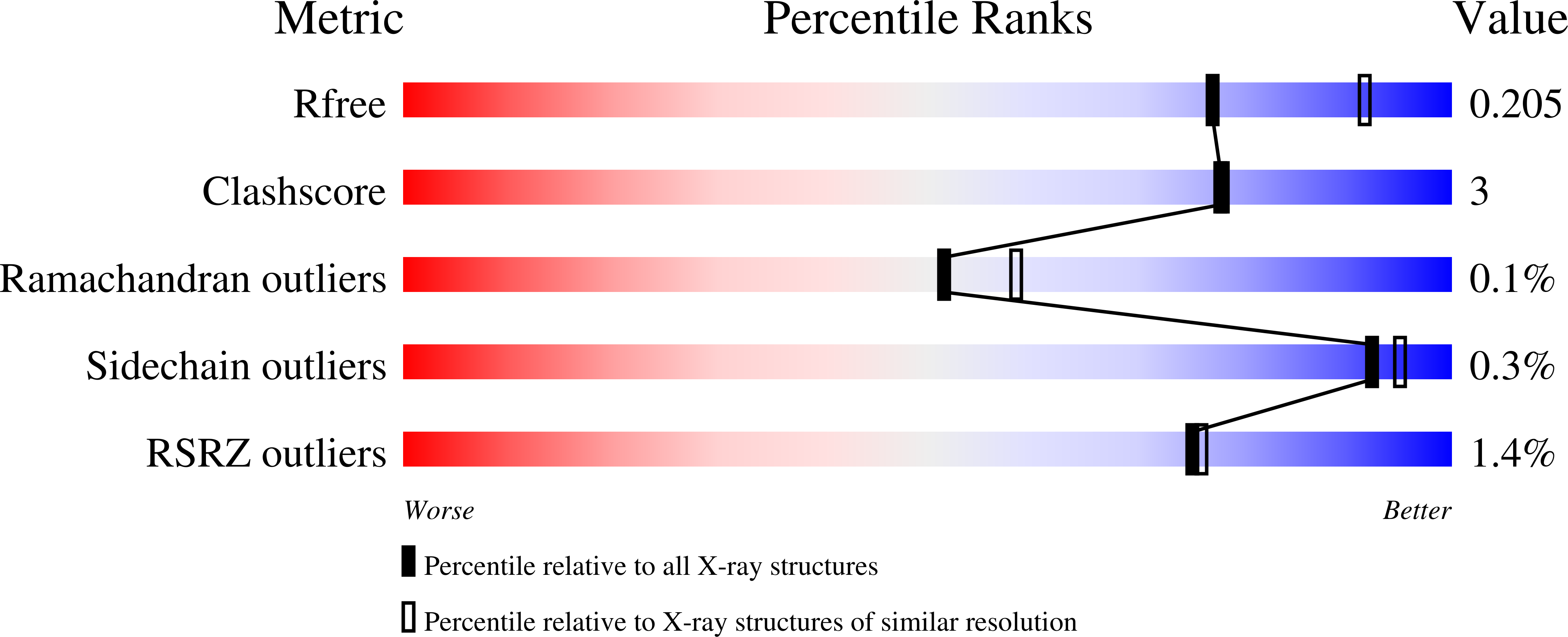
R-Value Free:
0.20
R-Value Work:
0.17
R-Value Observed:
0.17
Space Group:
P 21 21 21