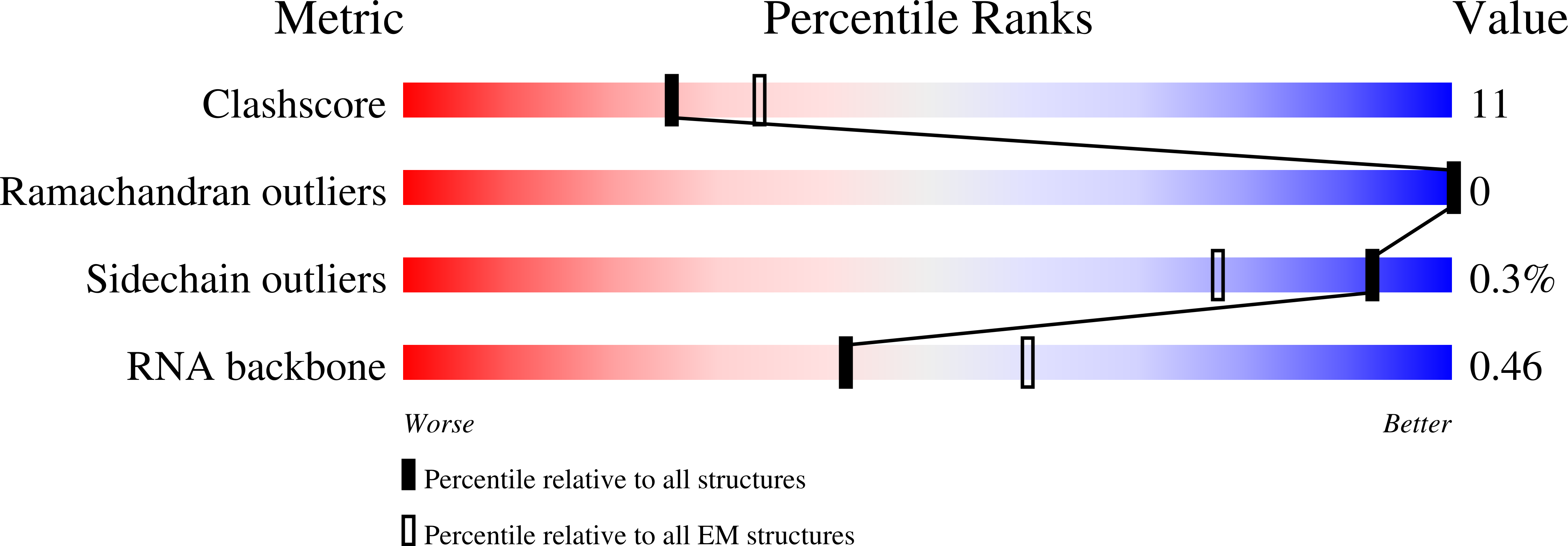
7UWS image
Entry Detail
PDB ID:
7UWS
EMDB ID:
Title:
Atomic model of the partial VSV nucleocapsid
Biological Source:
PDB Version:
Deposition Date:
2022-05-03
Release Date:
2022-09-28
Method Details:
Experimental Method:
Resolution:
3.47 Å
Aggregation State:
HELICAL ARRAY
Reconstruction Method:
HELICAL
Macromolecular Entities
Polymer Type:polypeptide(L)
Description:Nucleoprotein
Chain IDs:A, B, C, D, E, F, G
Chain Length:422
Number of Molecules:7
Biological Source:Vesicular stomatitis virus
Polymer Type:polyribonucleotide
Description:RNA
Chain IDs:H
Chain Length:381
Number of Molecules:1
Biological Source:Vesicular stomatitis virus
Polymer Type:polypeptide(L)
Description:Matrix protein
Chain IDs:I (auth: J), J (auth: K), K (auth: L), L (auth: M), M (auth: N), N (auth: O), O (auth: P), P (auth: Q), Q (auth: R), R (auth: S), S (auth: T), T (auth: U)
Chain Length:229
Number of Molecules:12
Biological Source:Vesicular stomatitis virus
Ligand Molecules
Primary Citation
Atomic model of vesicular stomatitis virus and mechanism of assembly.
Nat Commun 13 5980 5980 (2022)
PMID: 36216930 DOI: 10.1038/s41467-022-33664-4

Abstact

Like other negative-strand RNA viruses (NSVs) such as influenza and rabies, vesicular stomatitis virus (VSV) has a three-layered organization: a layer of matrix protein (M) resides between the glycoprotein (G)-studded membrane envelope and the nucleocapsid, which is composed of the nucleocapsid protein (N) and the encapsidated genomic RNA. Lack of in situ atomic structures of these viral components has limited mechanistic understanding of assembling the bullet-shaped virion. Here, by cryoEM and sub-particle reconstruction, we have determined the in situ structures of M and N inside VSV at 3.47 Å resolution. In the virion, N and M sites have a stoichiometry of 1:2. The in situ structures of both N and M differ from their crystal structures in their N-terminal segments and oligomerization loops. N-RNA, N-N, and N-M-M interactions govern the formation of the capsid. A double layer of M contributes to packaging of the helical nucleocapsid: the inner M (IM) joins neighboring turns of the N helix, while the outer M (OM) contacts G and the membrane envelope. The pseudo-crystalline organization of G is further mapped by cryoET. The mechanism of VSV assembly is delineated by the network interactions of these viral components.

Legend

Protein

Chemical

Disease

Primary Citation of related structures