
Deposition Date
2022-04-28
Release Date
2022-08-03
Last Version Date
2024-10-23
Entry Detail
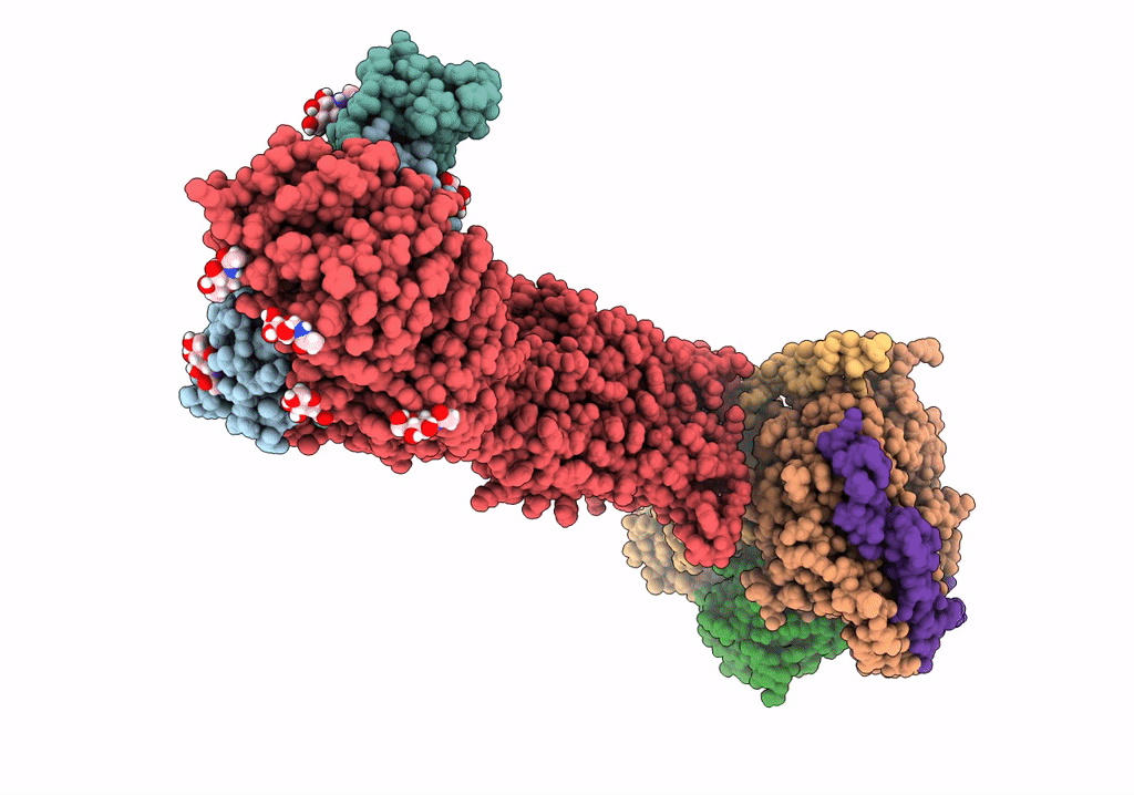
PDB ID:
7UTZ
Keywords:
Title:
Human thyrotropin analog TR1402 bound to human Thyrotropin receptor in complex with miniGs399 (composite structure)
Biological Source:
Source Organism(s):
Homo sapiens (Taxon ID: 9606)
Expression System(s):
Method Details:
Experimental Method:
Resolution:
2.40 Å
Aggregation State:
PARTICLE
Reconstruction Method:
SINGLE PARTICLE
