Deposition Date
2022-04-26
Release Date
2022-05-04
Last Version Date
2023-10-18
Entry Detail
PDB ID:
7UTC
Keywords:
Title:
Crystal structure of secondary alcohol dehydrogenases from the Thermoanaerobacter ethanolicus with NADP and transition-state analogue inhibitor DMSO
Biological Source:
Source Organism(s):
Thermoanaerobacter pseudethanolicus (Taxon ID: 496866)
Expression System(s):
Method Details:
Experimental Method:
Resolution:
1.85 Å
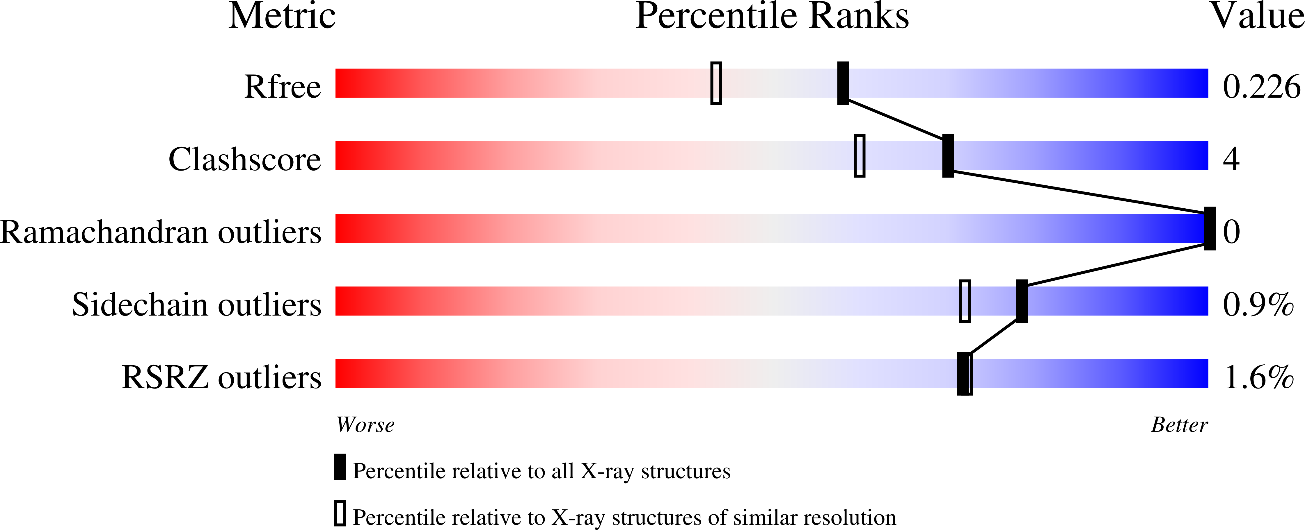
R-Value Free:
0.22
R-Value Work:
0.17
R-Value Observed:
0.17
Space Group:
P 21 21 21