Deposition Date
2022-04-26
Release Date
2022-09-21
Last Version Date
2024-10-23
Entry Detail
PDB ID:
7UT3
Keywords:
Title:
Crystal structure of complex of Fab, G10C with GalNAc-pNP
Biological Source:
Source Organism(s):
Mus musculus (Taxon ID: 10090)
Expression System(s):
Method Details:
Experimental Method:
Resolution:
3.00 Å
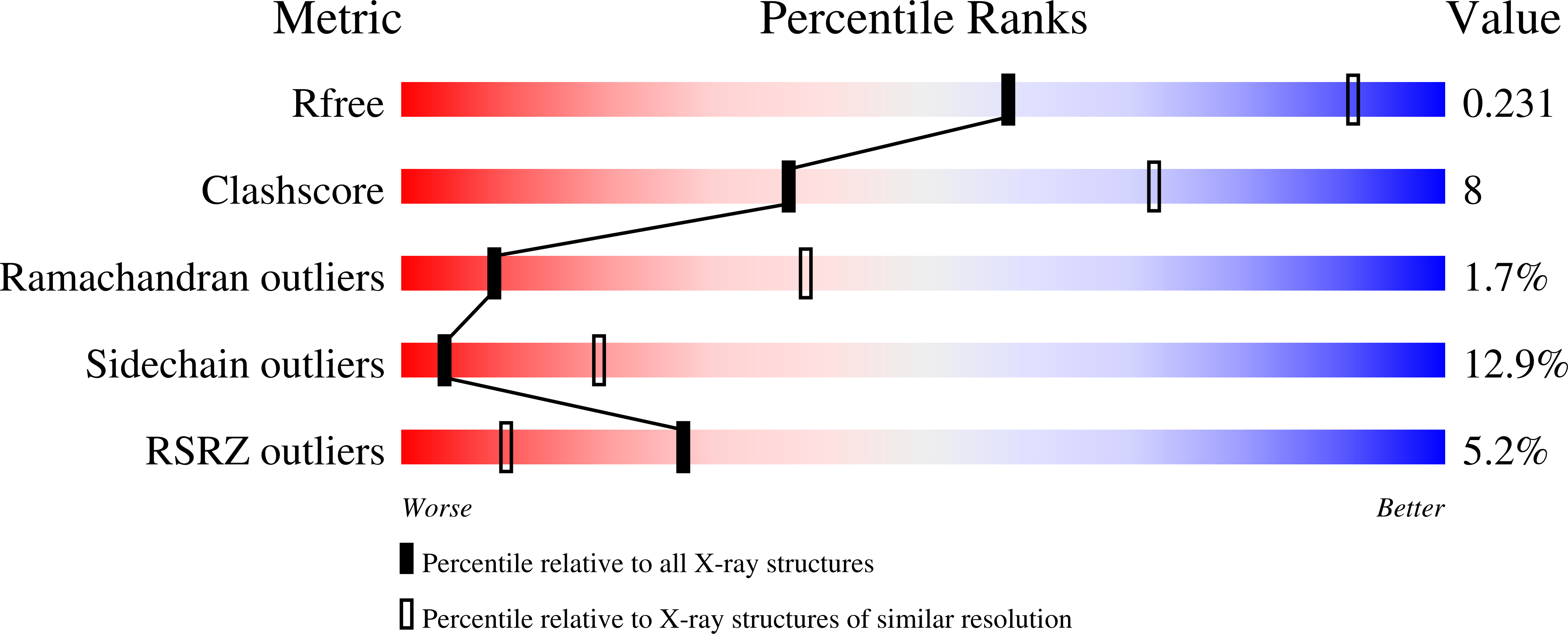
R-Value Free:
0.24
R-Value Work:
0.18
R-Value Observed:
0.18
Space Group:
P 61 2 2