7UPZ image
Deposition Date 2022-04-18
Release Date 2022-11-23
Last Version Date 2023-10-18
Entry Detail
PDB ID:
7UPZ
Title:
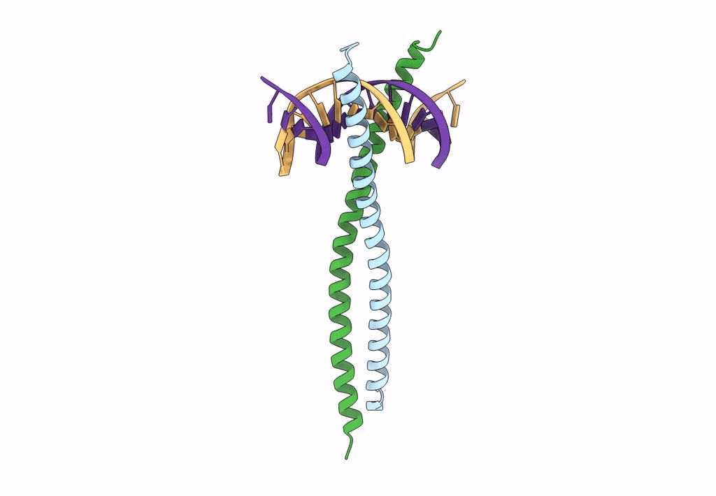
Structural basis for cell type specific DNA binding of C/EBPbeta: the case of cell cycle inhibitor p15INK4b promoter
Biological Source:
Source Organism:
Homo sapiens (Taxon ID: 9606)
Host Organism:
Method Details:
Experimental Method:
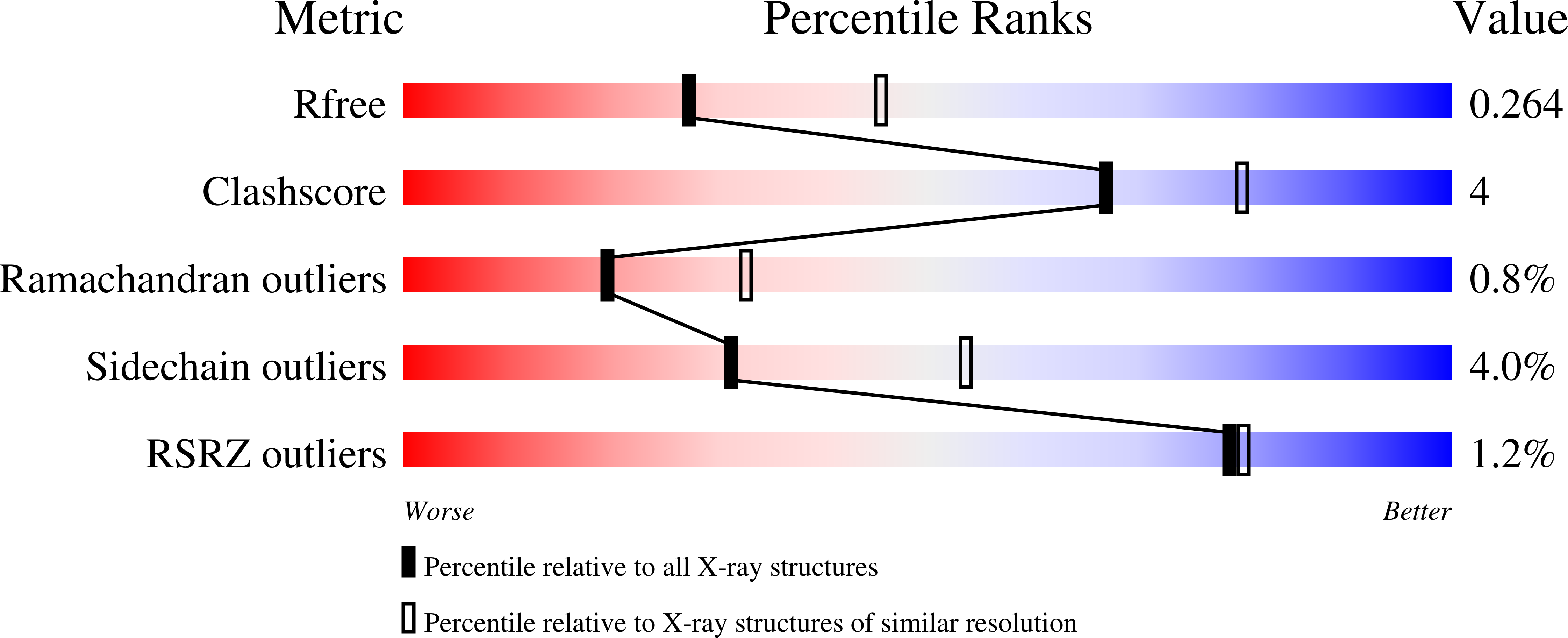
Resolution:
2.49 Å
R-Value Free:
0.26
R-Value Work:
0.21
R-Value Observed:
0.21
Space Group:
C 2 2 21
Macromolecular Entities
Polymer Type:polypeptide(L)
Molecule:CCAAT/enhancer-binding protein beta
Gene (Uniprot):CEBPB
Chain IDs:A, B
Chain Length:80
Number of Molecules:2
Biological Source:Homo sapiens
Polymer Type:polydeoxyribonucleotide
Molecule:DNA (5'-D(*AP*TP*TP*CP*TP*TP*AP*AP*GP*AP*AP*AP*GP*AP*CP*G)-3')
Chain IDs:C
Chain Length:16
Number of Molecules:1
Biological Source:Homo sapiens
Polymer Type:polydeoxyribonucleotide
Molecule:DNA (5'-D(*TP*CP*GP*TP*CP*TP*TP*TP*CP*TP*TP*AP*AP*GP*AP*A)-3')
Chain IDs:D
Chain Length:16
Number of Molecules:1
Biological Source:Homo sapiens
Primary Citation
Structural basis for cell type specific DNA binding of C/EBP beta : The case of cell cycle inhibitor p15INK4b promoter.
J.Struct.Biol. 214 107918 107918 (2022)
PMID: 36343842 DOI: 10.1016/j.jsb.2022.107918

Abstact

C/EBPβ is a key regulator of numerous cellular processes, but it can also contribute to tumorigenesis and viral diseases. It binds to specific DNA sequences (C/EBP sites) and interacts with other transcription factors to control expression of multiple eukaryotic genes in a tissue and cell-type dependent manner. A body of evidence has established that cell-type-specific regulatory information is contained in the local DNA sequence of the binding motif. In human epithelial cells, C/EBPβ is an essential cofactor for TGFβ signaling in the case of Smad2/3/4 and FoxO-dependent induction of the cell cycle inhibitor, p15INK4b. In the TGFβ-responsive region 2 of the p15INK4b promoter, the Smad binding site is flanked by a C/EBP site, CTTAA•GAAAG, which differs from the canonical, palindromic ATTGC•GCAAT motif. The X-ray crystal structure of C/EBPβ bound to the p15INK4b promoter fragment shows how GCGC-to-AAGA substitution generates changes in the intermolecular interactions in the protein-DNA interface that enhances C/EBPβ binding specificity, limits possible epigenetic regulation of the promoter, and generates a DNA element with a unique pattern of methyl groups in the major groove. Significantly, CT/GA dinucleotides located at the 5'ends of the double stranded element maintain local narrowing of the DNA minor groove width that is necessary for DNA recognition. Our results suggest that C/EBPβ would accept all forms of modified cytosine in the context of the CpT site. This contrasts with the effect on the consensus motif, where C/EBPβ binding is modestly increased by cytosine methylation, but substantially decreased by hydroxymethylation.

Legend

Protein

Chemical

Disease

Primary Citation of related structures