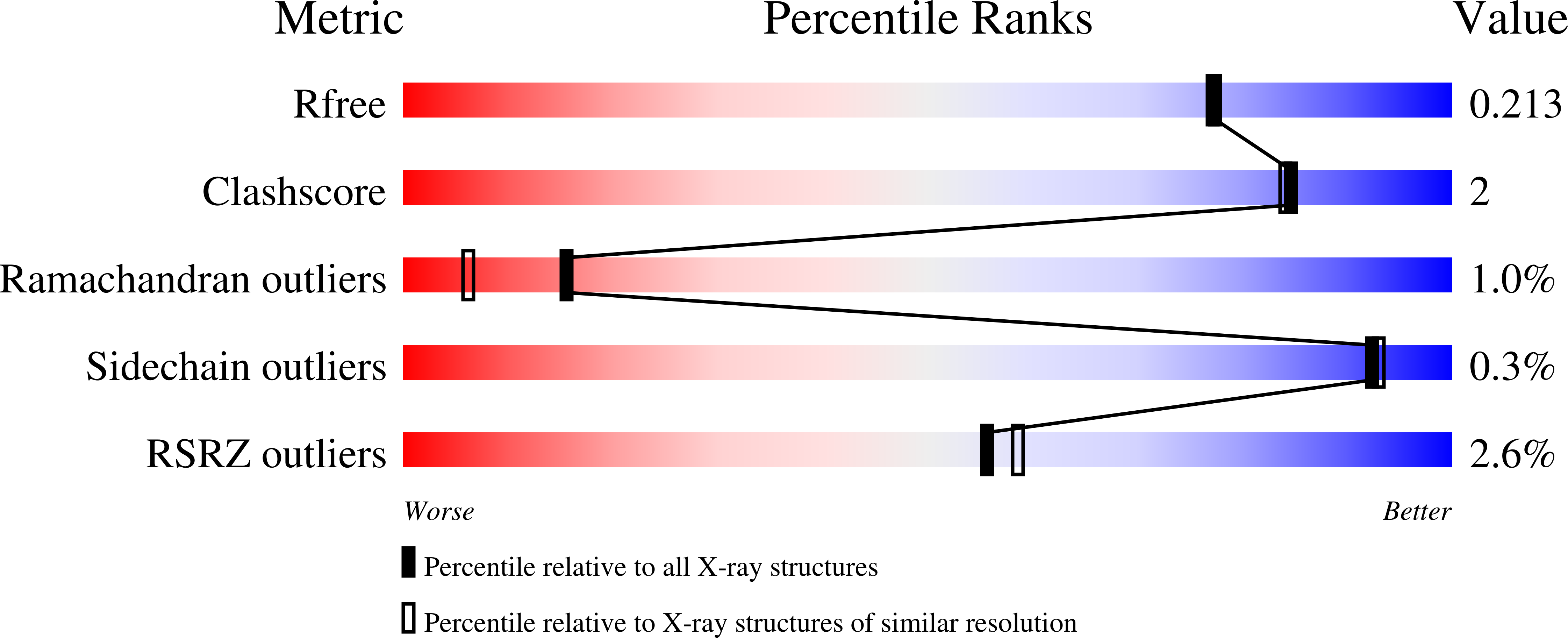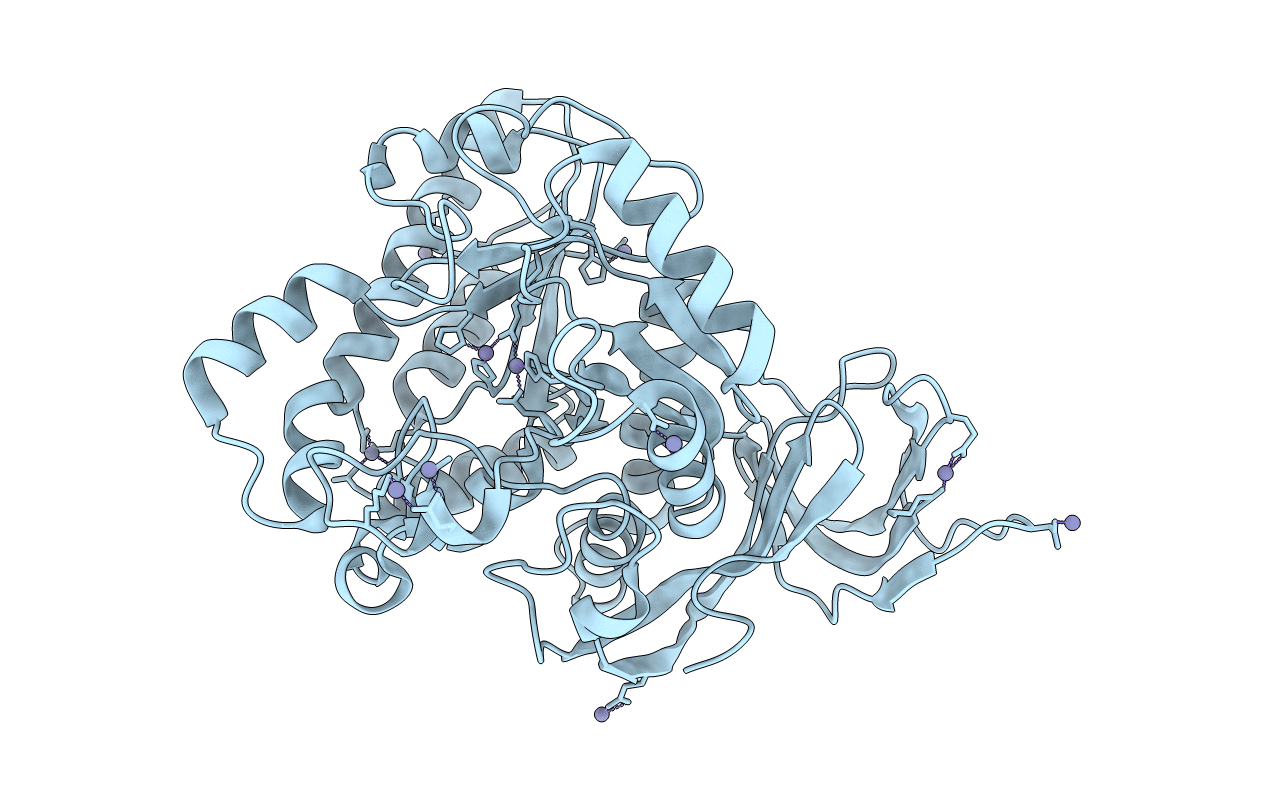
7UOF image
Deposition Date 2022-04-12
Release Date 2022-08-31
Last Version Date 2023-11-15
Entry Detail
PDB ID:
7UOF
Keywords:
Title:
Dihydroorotase from M. jannaschii
Biological Source:
Source Organism:
Host Organism:
Method Details:
Experimental Method:
Resolution:
1.90 Å
R-Value Free:
0.21
R-Value Work:
0.17
R-Value Observed:
0.17
Space Group:
P 32 2 1
Macromolecular Entities
Polymer Type:polypeptide(L)
Molecule:Dihydroorotase
Gene (Uniprot):pyrC
Chain IDs:A
Chain Length:423
Number of Molecules:1
Biological Source:Methanocaldococcus jannaschii
Modified Residue
Compound ID Chain ID Parent Comp ID Details 2D Image
KCX A LYS modified residue
Ligand Molecules
Primary Citation
Crystal structure of Methanococcus jannaschii dihydroorotase.
Proteins 91 91 98 (2023)
PMID: 35978488 DOI: 10.1002/prot.26412

Abstact

In this paper, we report the structural analysis of dihydroorotase (DHOase) from the hyperthermophilic and barophilic archaeon Methanococcus jannaschii. DHOase catalyzes the reversible cyclization of N-carbamoyl-l-aspartate to l-dihydroorotate in the third step of de novo pyrimidine biosynthesis. DHOases form a very diverse family of enzymes and have been classified into types and subtypes with structural similarities and differences among them. This is the first archaeal DHOase studied by x-ray diffraction. Its structure and comparison with known representatives of the other subtypes help define the structural features of the archaeal subtype. The M. jannaschii DHOase is found here to have traits from all subtypes. Contrary to expectations, it has a carboxylated lysine bridging the two Zn ions in the active site, and a long catalytic loop. It is a monomeric protein with a large β sandwich domain adjacent to the TIM barrel. Loop 5 is similar to bacterial type III and the C-terminal extension is long.

Legend

Protein

Chemical

Disease

Primary Citation of related structures