
Deposition Date
2022-04-07
Release Date
2023-10-11
Last Version Date
2023-10-11
Entry Detail
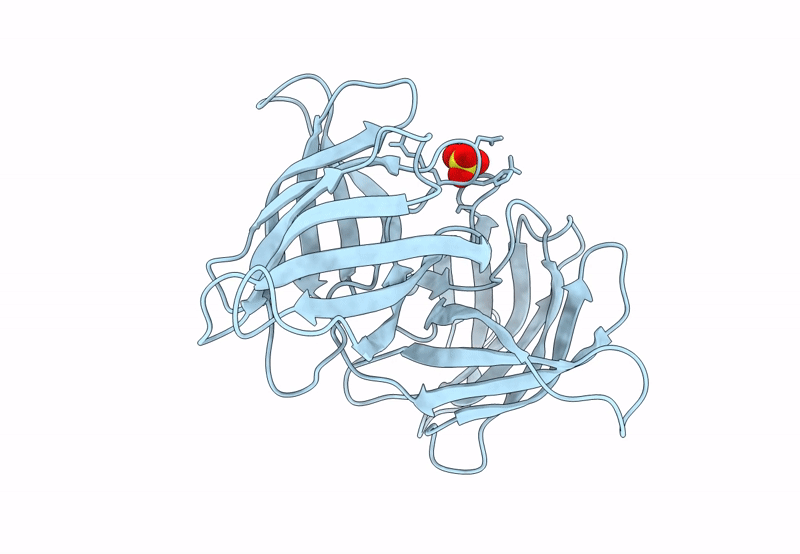
PDB ID:
7UMJ
Keywords:
Title:
Crystal structure of recombinant Solieria filiformis lectin (rSfL)
Biological Source:
Source Organism(s):
Solieria filiformis (Taxon ID: 31449)
Expression System(s):
Method Details:
Experimental Method:
Resolution:
1.88 Å
R-Value Free:
0.23
R-Value Work:
0.18
R-Value Observed:
0.18
Space Group:
P 2 21 21
