7UIS image
Deposition Date 2022-03-29
Release Date 2022-04-06
Last Version Date 2023-10-18
Entry Detail
PDB ID:
7UIS
Keywords:
Title:
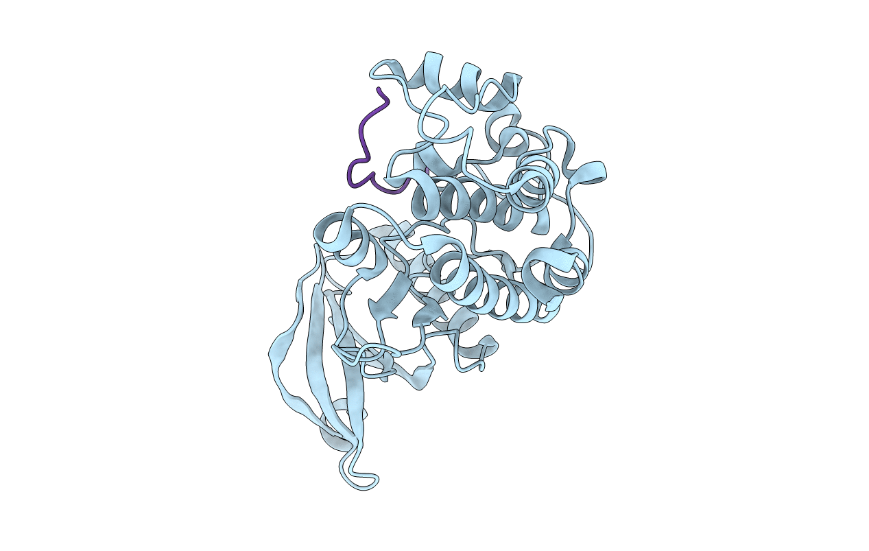
Cocrystal structure of human CaMKII-alpha (CAMK2A)kinase domain and GluN2B(S1303D)
Biological Source:
Source Organism:
Homo sapiens (Taxon ID: 9606)
Method Details:
Experimental Method:
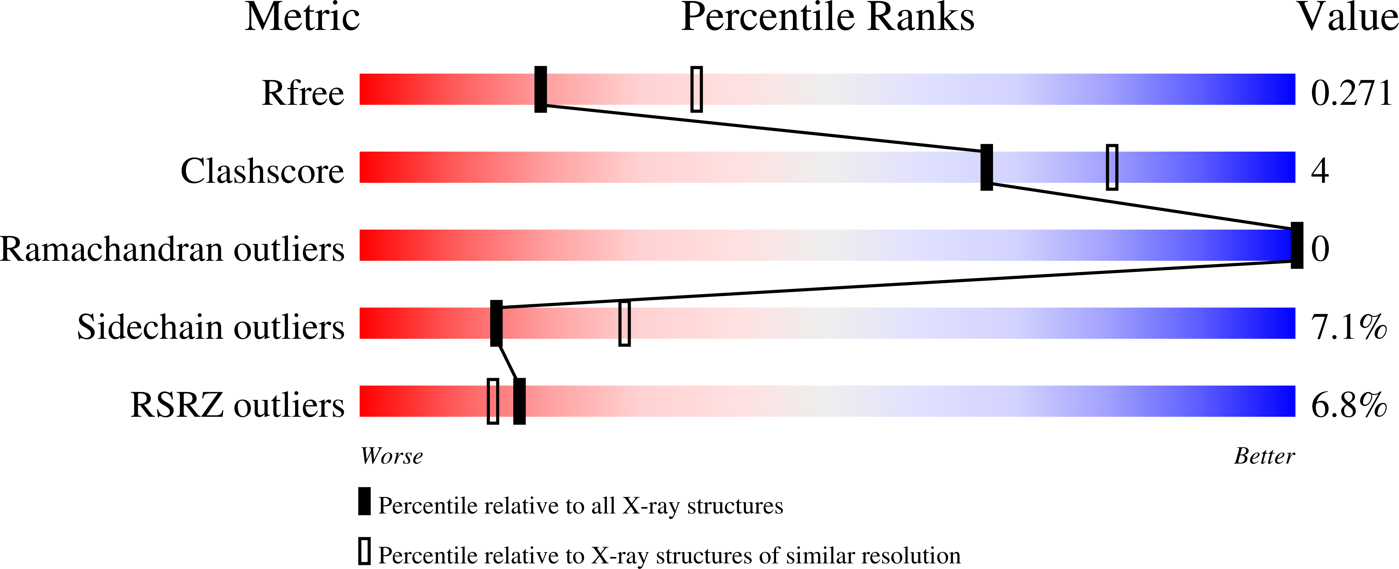
Resolution:
2.58 Å
R-Value Free:
0.26
R-Value Work:
0.21
R-Value Observed:
0.21
Space Group:
P 1 21 1
Macromolecular Entities
Structures with similar UniProt ID
Protein Blast
Polymer Type:polypeptide(L)
Molecule:Calcium/calmodulin-dependent protein kinase type II subunit alpha
Gene (Uniprot):CAMK2A
Mutagens:D135N, Q223K
Chain IDs:A
Chain Length:268
Number of Molecules:1
Biological Source:Homo sapiens
Structures with similar UniProt ID
Protein Blast
Polymer Type:polypeptide(L)
Molecule:Glutamate receptor ionotropic, NMDA 2B
Gene (Uniprot):GRIN2B
Mutagens:S1303D
Chain IDs:B
Chain Length:22
Number of Molecules:1
Biological Source:Homo sapiens
Ligand Molecules
Primary Citation
CaMKII binds both substrates and activators at the active site.
Cell Rep 40 111064 111064 (2022)
PMID: 35830796 DOI: 10.1016/j.celrep.2022.111064

Abstact

Ca2+/calmodulin-dependent protein kinase II (CaMKII) is a signaling protein required for long-term memory. When activated by Ca2+/CaM, it sustains activity even after the Ca2+ dissipates. In addition to the well-known autophosphorylation-mediated mechanism, interaction with specific binding partners also persistently activates CaMKII. A long-standing model invokes two distinct S and T sites. If an interactor binds at the T-site, then it will preclude autoinhibition and allow substrates to be phosphorylated at the S site. Here, we specifically test this model with X-ray crystallography, molecular dynamics simulations, and biochemistry. Our data are inconsistent with this model. Co-crystal structures of four different activators or substrates show that they all bind to a single continuous site across the kinase domain. We propose a mechanistic model where persistent CaMKII activity is facilitated by high-affinity binding partners that kinetically compete with autoinhibition by the regulatory segment to allow substrate phosphorylation.

Legend

Protein

Chemical

Disease

Primary Citation of related structures
Feedback Form
Name
Email
Institute
Feedback