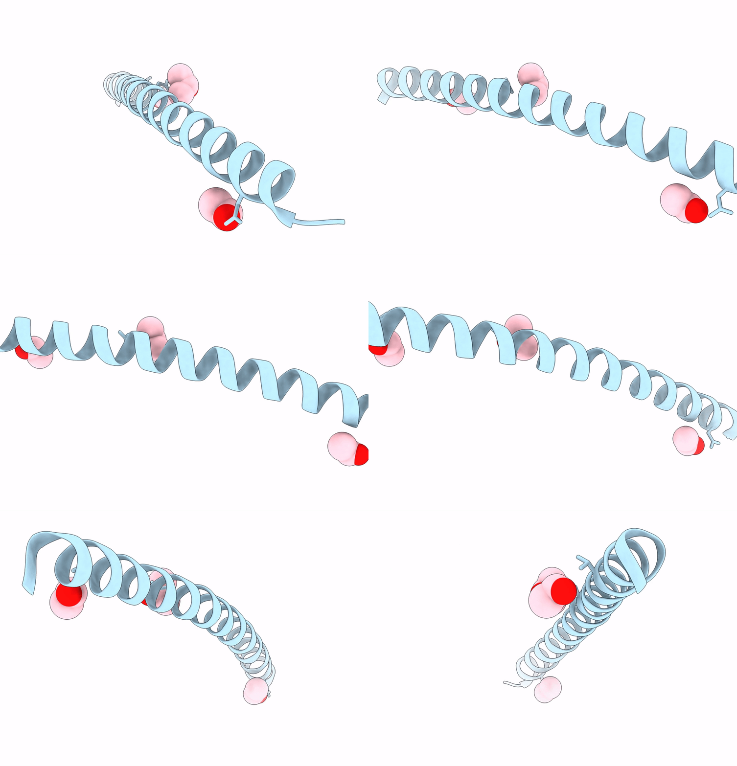
7UG2 image
Deposition Date 2022-03-23
Release Date 2023-02-15
Last Version Date 2023-10-25
Entry Detail
PDB ID:
7UG2
Keywords:
Title:
Crystal structure of the coiled-coil domain of TRIM75
Biological Source:
Source Organism:
Mus musculus (Taxon ID: 10090)
Method Details:
Experimental Method:
Resolution:
2.05 Å
R-Value Free:
0.25
R-Value Work:
0.21
Space Group:
I 41 2 2
Macromolecular Entities
Polymer Type:polypeptide(L)
Molecule:Tripartite motif-containing protein 75
Gene (Uniprot):Trim75
Chain IDs:A
Chain Length:59
Number of Molecules:1
Biological Source:Mus musculus
Primary Citation
Structural studies of the coiled-coil domain of TRIM75 reveal a tetramer architecture facilitating its E3 ligase complex.
Comput Struct Biotechnol J 20 4921 4929 (2022)
PMID: 36147661 DOI: 10.1016/j.csbj.2022.08.069

Abstact

Protein ubiquitination plays a vital role in controlling the degradation of intracellular proteins and in regulating cell signaling pathways. Functionally, E3 ubiquitin ligases control the transfer of ubiquitin to the target substrates. As a major family of ubiquitin E3 ligases, the structural assembly of RING E3 ligases required to exert their ubiquitin E3 ligase activity remains poorly defined. Here, we solved the crystal structure of the coiled-coil domain of TRIM75, a member of the RING E3 ligase family, which showed that two disulfide bonds stabilize two antiparallel dimers at a small crossing angle. This tetrameric conformation confers two close RING domains on the same side to form a dimer. Furthermore, this architecture allows the RING dimer to present ubiquitin to a substrate on the same side. Overall, this structure reveals a disulfide bond-mediated unique tetramer architecture and provides a tetrameric structural model through which E3 ligases exert their function.

Legend

Protein

Chemical

Disease

Primary Citation of related structures