
Deposition Date
2022-03-18
Release Date
2022-09-21
Last Version Date
2024-12-25
Entry Detail
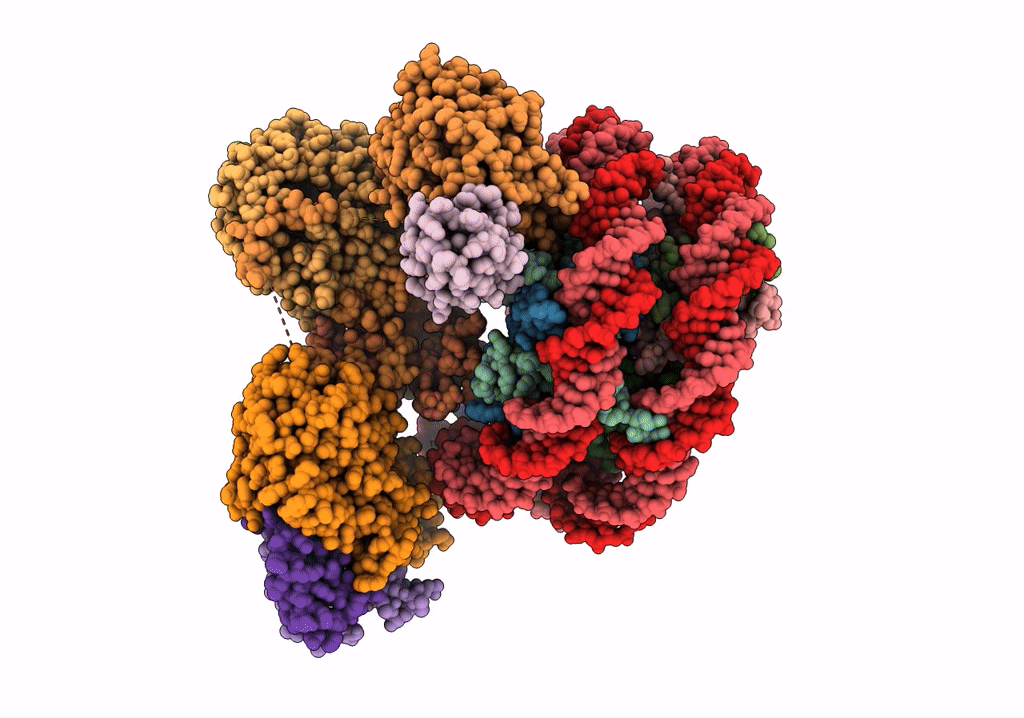
PDB ID:
7UD5
Keywords:
Title:
Complex between MLL1-WRAD and an H2B-ubiquitinated nucleosome
Biological Source:
Source Organism(s):
Xenopus laevis (Taxon ID: 8355)
synthetic construct (Taxon ID: 32630)
Homo sapiens (Taxon ID: 9606)
synthetic construct (Taxon ID: 32630)
Homo sapiens (Taxon ID: 9606)
Expression System(s):
Method Details:
Experimental Method:
Resolution:
4.25 Å
Aggregation State:
PARTICLE
Reconstruction Method:
SINGLE PARTICLE
