7UBB image
Deposition Date 2022-03-14
Release Date 2022-12-07
Last Version Date 2024-06-12
Entry Detail
PDB ID:
7UBB
Title:
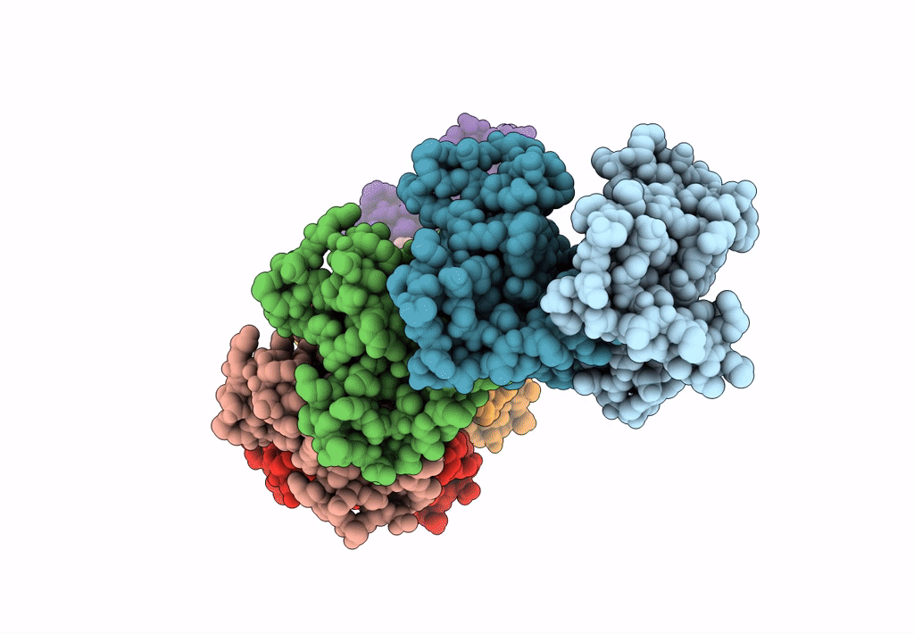
Structure of RecT protein from Listeria innoccua phage A118 in complex with 83-mer ssDNA
Biological Source:
Source Organism:
Host Organism:
Method Details:
Experimental Method:
Resolution:
4.50 Å
Aggregation State:
PARTICLE
Reconstruction Method:
SINGLE PARTICLE
Macromolecular Entities
Polymer Type:polypeptide(L)
Molecule:RecT
Gene (Uniprot):lin0085
Chain IDs:A (auth: C), B (auth: D), C (auth: E), D (auth: F), E (auth: G), F (auth: H), G (auth: I), H (auth: J)
Chain Length:274
Number of Molecules:8
Biological Source:Listeria innocua Clip11262
Ligand Molecules
Primary Citation
Structure of a RecT/Red beta family recombinase in complex with a duplex intermediate of DNA annealing.
Nat Commun 13 7855 7855 (2022)
PMID: 36543802 DOI: 10.1038/s41467-022-35572-z

Abstact

Some bacteriophage encode a recombinase that catalyzes single-stranded DNA annealing (SSA). These proteins are apparently related to RAD52, the primary human SSA protein. The best studied protein, Redβ from bacteriophage λ, binds weakly to ssDNA, not at all to dsDNA, but tightly to a duplex intermediate of annealing formed when two complementary DNA strands are added to the protein sequentially. We used single particle cryo-electron microscopy (cryo-EM) to determine a 3.4 Å structure of a Redβ homolog from a prophage of Listeria innocua in complex with two complementary 83mer oligonucleotides. The structure reveals a helical protein filament bound to a DNA duplex that is highly extended and unwound. Native mass spectrometry confirms that the complex seen by cryo-EM is the predominant species in solution. The protein shares a common core fold with RAD52 and a similar mode of ssDNA-binding. These data provide insights into the mechanism of protein-catalyzed SSA.

Legend

Protein

Chemical

Disease

Primary Citation of related structures