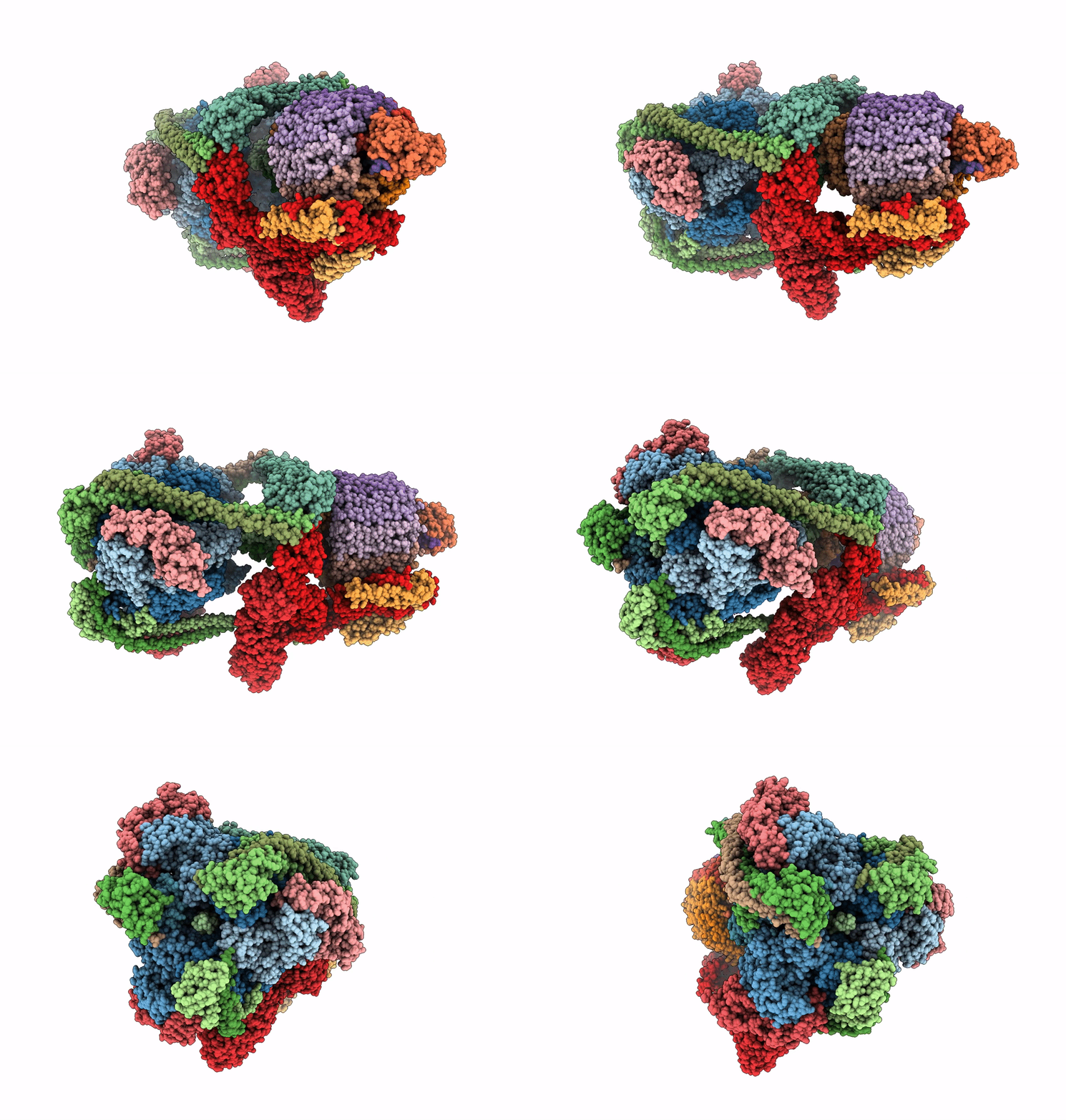
7U8P image
Entry Detail
PDB ID:
7U8P
EMDB ID:
Title:
Structure of porcine kidney V-ATPase with SidK, Rotary State 1
Biological Source:
Host Organism:
PDB Version:
Deposition Date:
2022-03-09
Release Date:
2022-07-06
Method Details:
Experimental Method:
Resolution:
3.70 Å
Aggregation State:
PARTICLE
Reconstruction Method:
SINGLE PARTICLE
Macromolecular Entities
Polymer Type:polypeptide(L)
Description:V-type proton ATPase catalytic subunit A
Chain IDs:A, B, C
Chain Length:617
Number of Molecules:3
Biological Source:Sus scrofa
Polymer Type:polypeptide(L)
Description:Vacuolar proton pump subunit B
Chain IDs:D, E, F
Chain Length:351
Number of Molecules:3
Biological Source:Sus scrofa
Polymer Type:polypeptide(L)
Description:V-type proton ATPase subunit C
Chain IDs:G
Chain Length:155
Number of Molecules:1
Biological Source:Sus scrofa
Polymer Type:polypeptide(L)
Description:V-type proton ATPase subunit D
Chain IDs:H
Chain Length:247
Number of Molecules:1
Biological Source:Sus scrofa
Polymer Type:polypeptide(L)
Description:V-type proton ATPase subunit E 1
Chain IDs:I, J, K
Chain Length:226
Number of Molecules:3
Biological Source:Sus scrofa
Polymer Type:polypeptide(L)
Description:V-type proton ATPase subunit F
Chain IDs:L
Chain Length:119
Number of Molecules:1
Biological Source:Sus scrofa
Polymer Type:polypeptide(L)
Description:V-type proton ATPase subunit G
Chain IDs:M, N, O
Chain Length:118
Number of Molecules:3
Biological Source:Sus scrofa
Polymer Type:polypeptide(L)
Description:Bacterial effector protein SidK
Chain IDs:P (auth: Q), Q (auth: R), R (auth: S)
Chain Length:337
Number of Molecules:3
Biological Source:Legionella pneumophila
Polymer Type:polypeptide(L)
Description:V-type proton ATPase subunit H
Chain IDs:S (auth: T)
Chain Length:483
Number of Molecules:1
Biological Source:Sus scrofa
Polymer Type:polypeptide(L)
Description:V-type proton ATPase subunit a
Chain IDs:T (auth: a)
Chain Length:617
Number of Molecules:1
Biological Source:Sus scrofa
Polymer Type:polypeptide(L)
Description:V-type proton ATPase 21 kDa proteolipid subunit isoform 1
Chain IDs:U (auth: b)
Chain Length:205
Number of Molecules:1
Biological Source:Sus scrofa
Polymer Type:polypeptide(L)
Description:ATPase H+ transporting accessory protein 1
Chain IDs:V (auth: c)
Chain Length:469
Number of Molecules:1
Biological Source:Sus scrofa
Polymer Type:polypeptide(L)
Description:V-type proton ATPase subunit
Chain IDs:W (auth: d)
Chain Length:351
Number of Molecules:1
Biological Source:Sus scrofa
Polymer Type:polypeptide(L)
Description:V-type proton ATPase subunit
Chain IDs:X (auth: e)
Chain Length:515
Number of Molecules:1
Biological Source:Sus scrofa
Polymer Type:polypeptide(L)
Description:Ribonuclease kappa
Chain IDs:Y (auth: f)
Chain Length:515
Number of Molecules:1
Biological Source:Sus scrofa
Polymer Type:polypeptide(L)
Description:V-type proton ATPase proteolipid subunit
Chain IDs:Z (auth: g), AA (auth: h), BA (auth: i), CA (auth: j), DA (auth: k), EA (auth: l), FA (auth: m), GA (auth: n), HA (auth: o)
Chain Length:155
Number of Molecules:9
Biological Source:Sus scrofa
Polymer Type:polypeptide(L)
Description:ATPase H(+)-transporting lysosomal accessory protein 2
Chain IDs:IA (auth: p)
Chain Length:351
Number of Molecules:1
Biological Source:Sus scrofa
Ligand Molecules
Primary Citation
CryoEM of endogenous mammalian V-ATPase interacting with the TLDc protein mEAK-7.
Life Sci Alliance 5 ? ? (2022)
PMID: 35794005 DOI: 10.26508/lsa.202201527

Abstact

V-ATPases are rotary proton pumps that serve as signaling hubs with numerous protein binding partners. CryoEM with exhaustive focused classification allowed detection of endogenous proteins associated with porcine kidney V-ATPase. An extra C subunit was found in ∼3% of complexes, whereas ∼1.6% of complexes bound mEAK-7, a protein with proposed roles in dauer formation in nematodes and mTOR signaling in mammals. High-resolution cryoEM of porcine kidney V-ATPase with recombinant mEAK-7 showed that mEAK-7's TLDc domain interacts with V-ATPase's stator, whereas its C-terminal α helix binds V-ATPase's rotor. This crosslink would be expected to inhibit rotary catalysis. However, unlike the yeast TLDc protein Oxr1p, exogenous mEAK-7 does not inhibit V-ATPase and mEAK-7 overexpression in cells does not alter lysosomal or phagosomal pH. Instead, cryoEM suggests that the mEAK-7:V-ATPase interaction is disrupted by ATP-induced rotation of the rotor. Comparison of Oxr1p and mEAK-7 binding explains this difference. These results show that V-ATPase binding by TLDc domain proteins can lead to effects ranging from strong inhibition to formation of labile interactions that are sensitive to the enzyme's activity.

Legend

Protein

Chemical

Disease

Primary Citation of related structures