7U6V image
Entry Detail
PDB ID:
7U6V
EMDB ID:
Keywords:
Title:
Cryo-EM structure of Shiga toxin 2 in complex with the native ribosomal P-stalk
Biological Source:
PDB Version:
Deposition Date:
2022-03-06
Release Date:
2023-01-11
Method Details:
Experimental Method:
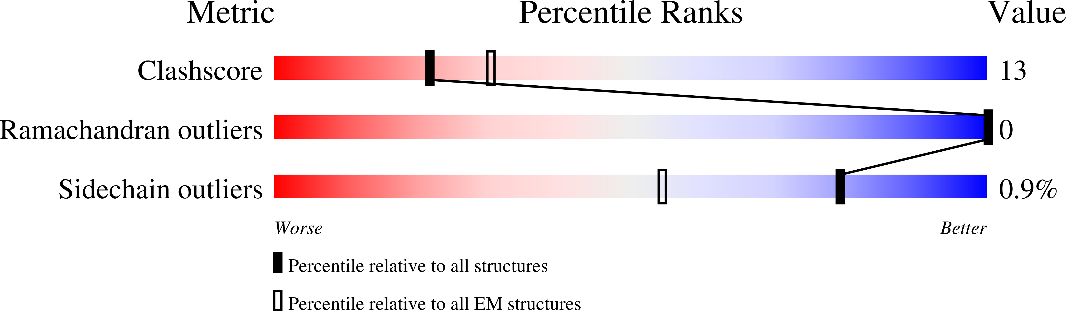
Resolution:
4.10 Å
Aggregation State:
PARTICLE
Reconstruction Method:
SINGLE PARTICLE
Macromolecular Entities
Polymer Type:polypeptide(L)
Description:Shiga toxin 2a subunit A (Stx2A)
Chain IDs:A
Chain Length:297
Number of Molecules:1
Biological Source:Shigella dysenteriae
Polymer Type:polypeptide(L)
Description:Shiga toxin 2a subunit B (Stx2B)
Chain IDs:B, C, D, E, F
Chain Length:70
Number of Molecules:5
Biological Source:Shigella dysenteriae
Polymer Type:polypeptide(L)
Description:C-terminal domain (CTD) from the Ribosomal P-stalk
Chain IDs:G (auth: P)
Chain Length:6
Number of Molecules:1
Biological Source:Saccharomyces cerevisiae
Primary Citation
Cryo-EM structure of Shiga toxin 2 in complex with the native ribosomal P-stalk reveals residues involved in the binding interaction.
J.Biol.Chem. 299 102795 102795 (2022)
PMID: 36528064 DOI: 10.1016/j.jbc.2022.102795

Abstact

Shiga toxin 2a (Stx2a) is the virulence factor of enterohemorrhagic Escherichia coli. The catalytic A1 subunit of Stx2a (Stx2A1) interacts with the ribosomal P-stalk for loading onto the ribosome and depurination of the sarcin-ricin loop, which halts protein synthesis. Because of the intrinsic flexibility of the P-stalk, a structure of the Stx2a-P-stalk complex is currently unknown. We demonstrated that the native P-stalk pentamer binds to Stx2a with nanomolar affinity, and we employed cryo-EM to determine a structure of the 72 kDa Stx2a complexed with the P-stalk. The structure identifies Stx2A1 residues involved in binding and reveals that Stx2a is anchored to the P-stalk via only the last six amino acids from the C-terminal domain of a single P-protein. For the first time, the cryo-EM structure shows the loop connecting Stx2A1 and Stx2A2, which is critical for activation of the toxin. Our principal component analysis of the cryo-EM data reveals the intrinsic dynamics of the Stx2a-P-stalk interaction, including conformational changes in the P-stalk binding site occurring upon complex formation. Our computational analysis unveils the propensity for structural rearrangements within the C-terminal domain, with its C-terminal six amino acids transitioning from a random coil to an α-helix upon binding to Stx2a. In conclusion, our cryo-EM structure sheds new light into the dynamics of the Stx2a-P-stalk interaction and indicates that the binding interface between Stx2a and the P-stalk is the potential target for drug discovery.

Legend

Protein

Chemical

Disease

Primary Citation of related structures