Deposition Date
2022-02-11
Release Date
2022-05-18
Last Version Date
2023-10-18
Entry Detail
PDB ID:
7TYB
Keywords:
Title:
Salicylate Adenylate PchD from Pseudomonas aeruginosa containing salicyl-AMS
Biological Source:
Source Organism(s):
Pseudomonas aeruginosa PAO1 (Taxon ID: 208964)
Expression System(s):
Method Details:
Experimental Method:
Resolution:
2.11 Å
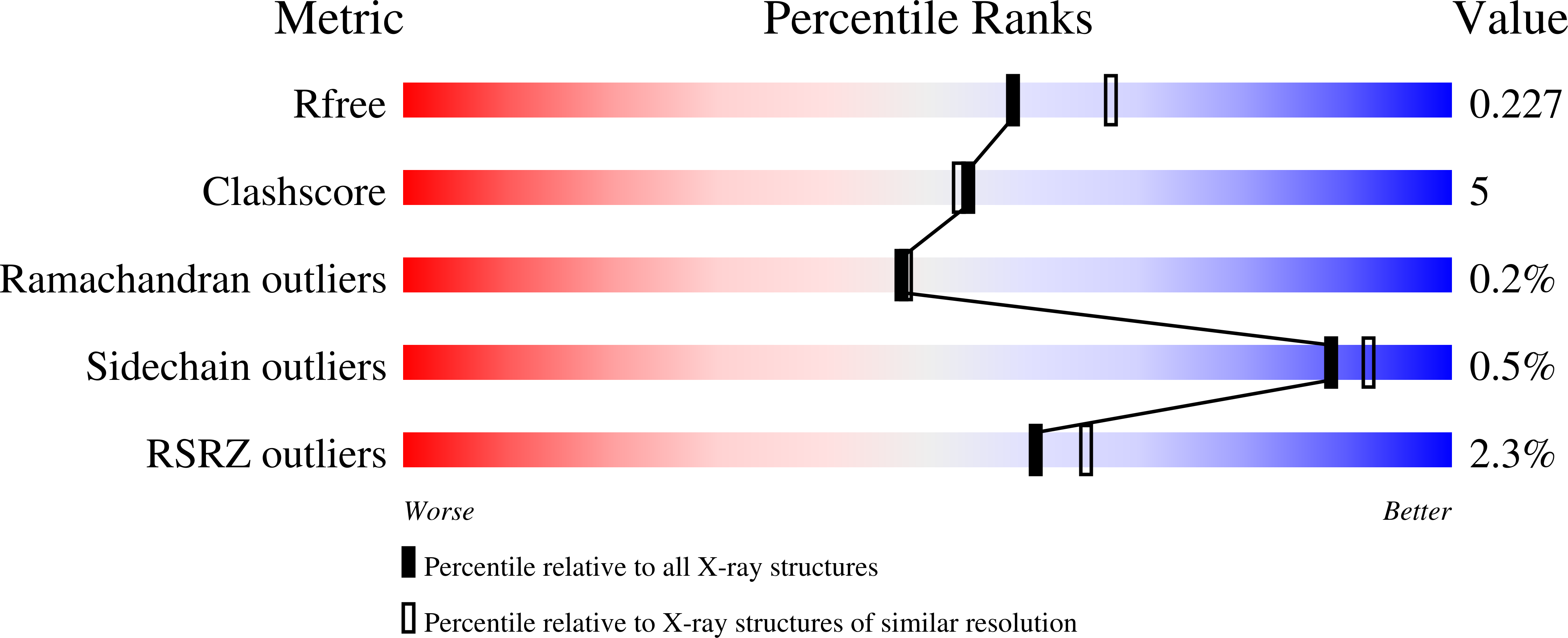
R-Value Free:
0.22
R-Value Work:
0.16
R-Value Observed:
0.17
Space Group:
C 1 2 1