
Deposition Date
2022-02-09
Release Date
2022-03-09
Last Version Date
2023-10-18
Entry Detail
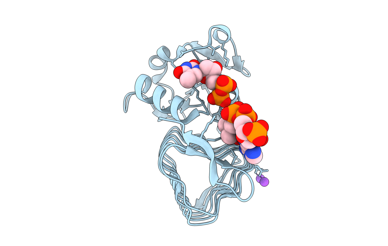
PDB ID:
7TXQ
Keywords:
Title:
x-ray structure of the VioB N-acetyltransferase from Acinetobacter baumannii in the present of TDP and Acetyl-CoenzymeA
Biological Source:
Source Organism(s):
Acinetobacter baumannii (Taxon ID: 470)
Expression System(s):
Method Details:
Experimental Method:
Resolution:
1.65 Å
R-Value Free:
0.20
R-Value Work:
0.17
R-Value Observed:
0.17
Space Group:
H 3
