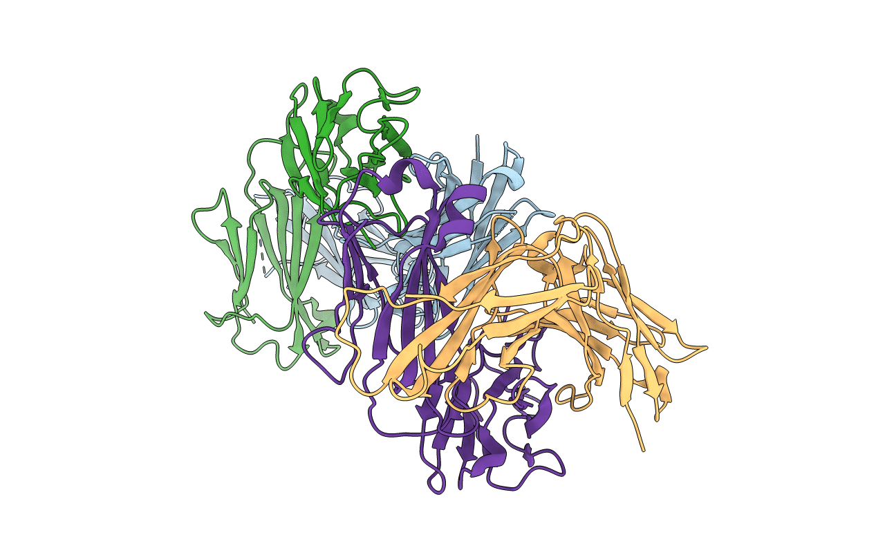
7TUH image
Entry Detail
PDB ID:
7TUH
Keywords:
Title:
Crystal structure of anti-tapasin PaSta2-Fab
Biological Source:
Source Organism:
Host Organism:
PDB Version:
Deposition Date:
2022-02-02
Release Date:
2022-09-07
Method Details:
Experimental Method:
Resolution:
2.30 Å
R-Value Free:
0.23
R-Value Work:
0.21
R-Value Observed:
0.22
Space Group:
P 1 21 1
Macromolecular Entities
Polymer Type:polypeptide(L)
Description:PaSta2 Fab heavy chain
Chain IDs:A (auth: H), C (auth: A)
Chain Length:230
Number of Molecules:2
Biological Source:Mus musculus
Polymer Type:polypeptide(L)
Description:PaSta2 Fab kappa light chain
Chain IDs:B (auth: L), D (auth: B)
Chain Length:220
Number of Molecules:2
Biological Source:Mus musculus
Primary Citation
Structural mechanism of tapasin-mediated MHC-I peptide loading in antigen presentation.
Nat Commun 13 5470 5470 (2022)
PMID: 36115831 DOI: 10.1038/s41467-022-33153-8

Abstact

Loading of MHC-I molecules with peptide by the catalytic chaperone tapasin in the peptide loading complex plays a critical role in antigen presentation and immune recognition. Mechanistic insight has been hampered by the lack of detailed structural information concerning tapasin-MHC-I. We present here crystal structures of human tapasin complexed with the MHC-I molecule HLA-B*44:05, and with each of two anti-tapasin antibodies. The tapasin-stabilized peptide-receptive state of HLA-B*44:05 is characterized by distortion of the peptide binding groove and destabilization of the β2-microglobulin interaction, leading to release of peptide. Movements of the membrane proximal Ig-like domains of tapasin, HLA-B*44:05, and β2-microglobulin accompany the transition to a peptide-receptive state. Together this ensemble of crystal structures provides insights into a distinct mechanism of tapasin-mediated peptide exchange.

Legend

Protein

Chemical

Disease

Primary Citation of related structures