Deposition Date
2022-01-31
Release Date
2022-11-09
Last Version Date
2023-11-15
Entry Detail
PDB ID:
7TSR
Keywords:
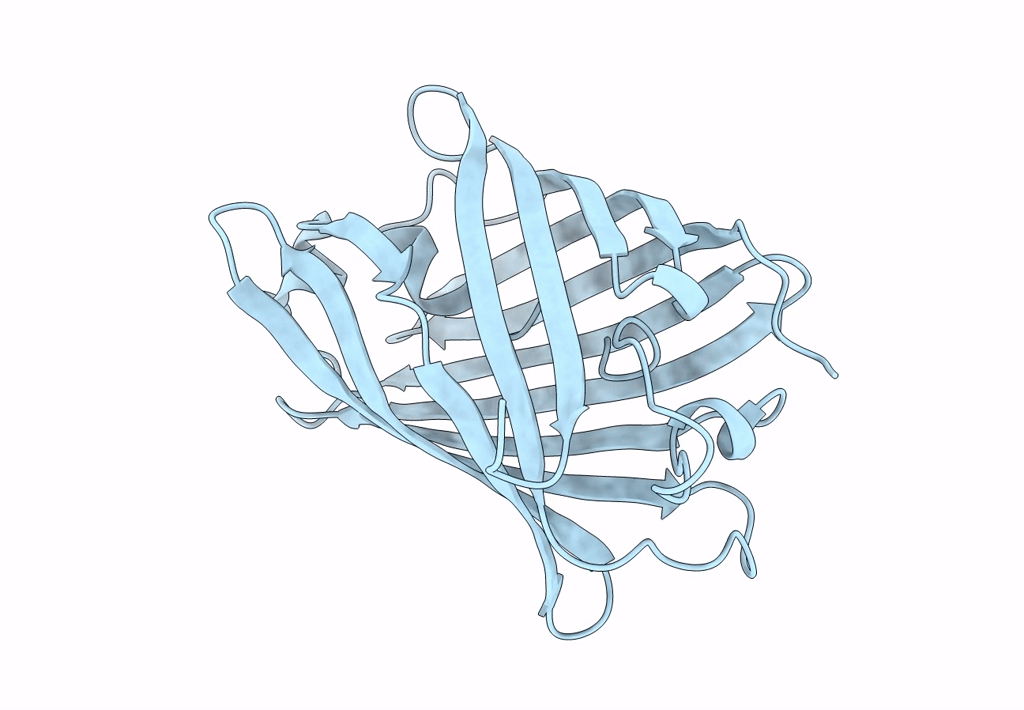
Title:
Room temperature rsEospa Cis-state structure at pH 8.4
Biological Source:
Source Organism(s):
Lobophyllia hemprichii (Taxon ID: 46758)
Expression System(s):
Method Details:
Experimental Method:
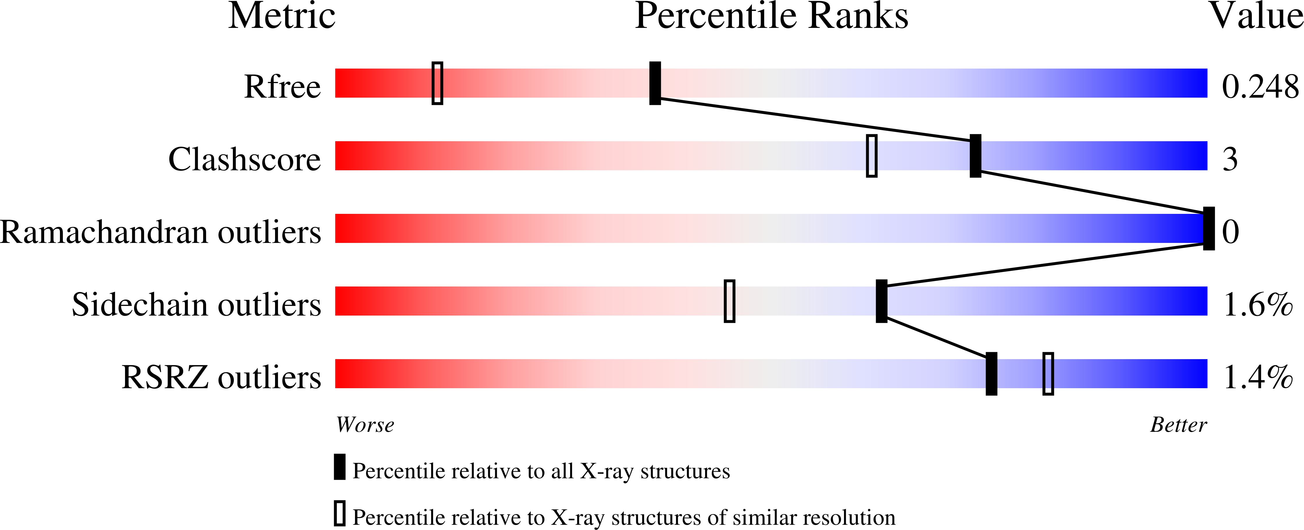
Resolution:
1.75 Å
R-Value Free:
0.24
R-Value Work:
0.21
Space Group:
P 21 21 21