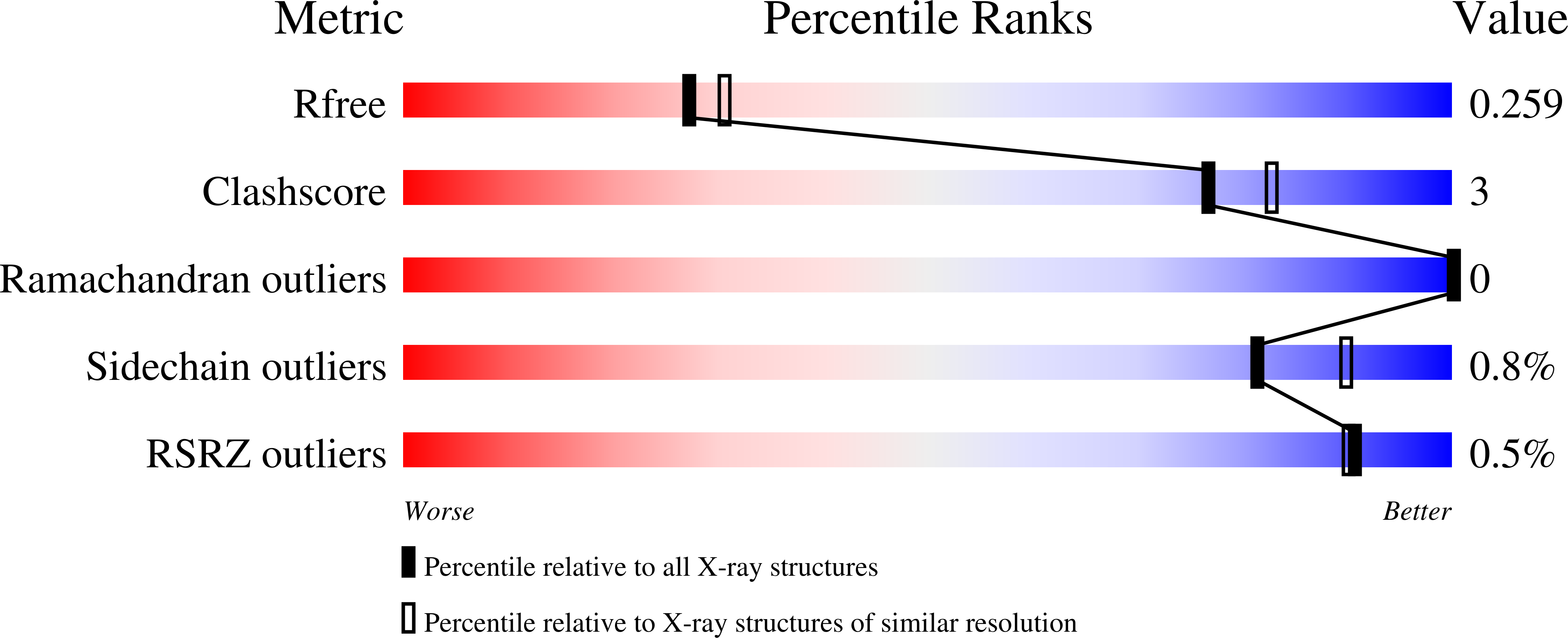
7TPU image
Entry Detail
PDB ID:
7TPU
Keywords:
Title:
Crystal structure of a chitinase-modifying protein from Fusarium vanettenii (Fvan-cmp)
Biological Source:
Source Organism:
Host Organism:
PDB Version:
Deposition Date:
2022-01-26
Release Date:
2023-02-15
Method Details:
Experimental Method:
Resolution:
2.19 Å
R-Value Free:
0.25
R-Value Work:
0.19
Space Group:
P 21 21 21
Macromolecular Entities
Polymer Type:polypeptide(L)
Description:Beta-lactamase domain-containing protein
Chain IDs:A
Chain Length:616
Number of Molecules:1
Biological Source:Fusarium vanettenii
Primary Citation
Crystal structure of a polyglycine hydrolase determined using a RoseTTAFold model.
Acta Crystallogr D Struct Biol 79 168 176 (2023)
PMID: 36762862 DOI: 10.1107/S2059798323000311

Abstact

Polyglycine hydrolases (PGHs) are secreted fungal proteases that cleave the polyglycine linker of Zea mays ChitA, a defensive chitinase, thus overcoming one mechanism of plant resistance to infection. Despite their importance in agriculture, there has been no previous structural characterization of this family of proteases. The objective of this research was to investigate the proteolytic mechanism and other characteristics by structural and biochemical means. Here, the first atomic structure of a polyglycine hydrolase was identified. It was solved by X-ray crystallography using a RoseTTAFold model, taking advantage of recent technical advances in structure prediction. PGHs are composed of two domains: the N- and C-domains. The N-domain is a novel tertiary fold with an as-yet unknown function that is found across all kingdoms of life. The C-domain shares structural similarities with class C β-lactamases, including a common catalytic nucleophilic serine. In addition to insights into the PGH family and its relationship to β-lactamases, the results demonstrate the power of complementing experimental structure determination with new computational techniques.

Legend

Protein

Chemical

Disease

Primary Citation of related structures