7TOH image
Entry Detail
PDB ID:
7TOH
Keywords:
Title:
Crystal structure of carbohydrate esterase PbeAcXE, in complex with MeGlcpA-Xylp
Biological Source:
PDB Version:
Deposition Date:
2022-01-24
Release Date:
2022-04-13
Method Details:
Experimental Method:
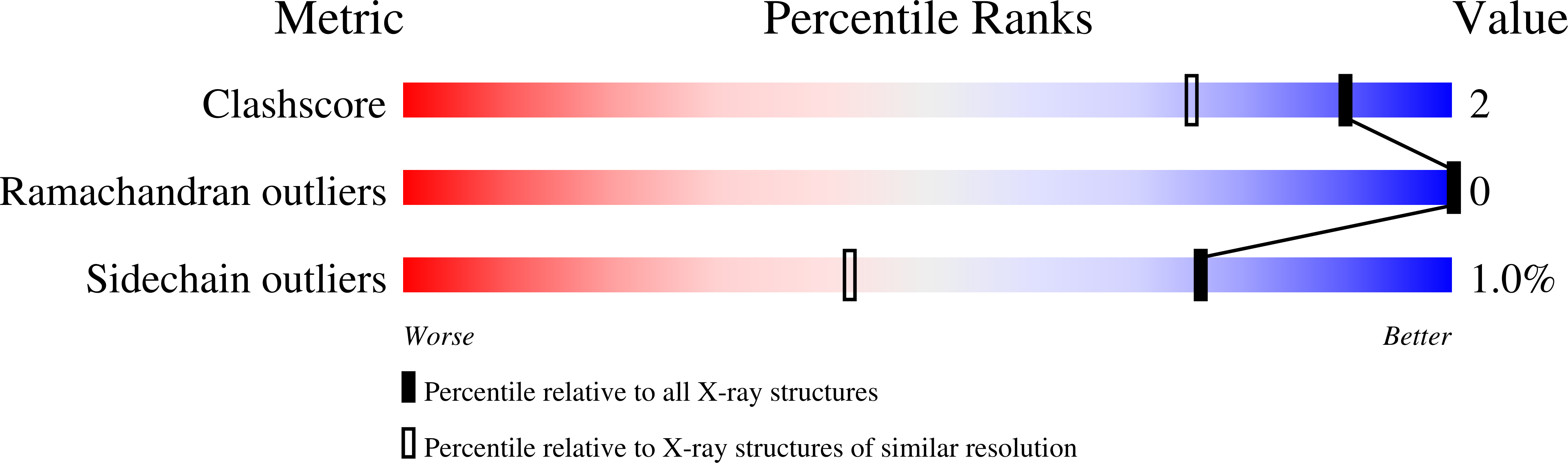
Resolution:
1.26 Å
R-Value Free:
0.16
R-Value Work:
0.13
R-Value Observed:
0.13
Space Group:
P 1 21 1
Macromolecular Entities
Polymer Type:polypeptide(L)
Description:SGNH hydrolase
Chain IDs:A
Chain Length:386
Number of Molecules:1
Biological Source:Prolixibacter bellariivorans
Modified Residue
Compound ID Chain ID Parent Comp ID Details 2D Image
MSE A MET modified residue
Primary Citation
Elucidating Sequence and Structural Determinants of Carbohydrate Esterases for Complete Deacetylation of Substituted Xylans.
Molecules 27 ? ? (2022)
PMID: 35566004 DOI: 10.3390/molecules27092655

Abstact

Acetylated glucuronoxylan is one of the most common types of hemicellulose in nature. The structure is formed by a β-(1→4)-linked D-xylopyranosyl (Xylp) backbone that can be substituted with an acetyl group at O-2 and O-3 positions, and α-(1→2)-linked 4-O-methylglucopyranosyluronic acid (MeGlcpA). Acetyl xylan esterases (AcXE) that target mono- or doubly acetylated Xylp are well characterized; however, the previously studied AcXE from Flavobacterium johnsoniae (FjoAcXE) was the first to remove the acetyl group from 2-O-MeGlcpA-3-O-acetyl-substituted Xylp units, yet structural characteristics of these enzymes remain unspecified. Here, six homologs of FjoAcXE were produced and three crystal structures of the enzymes were solved. Two of them are complex structures, one with bound MeGlcpA and another with acetate. All homologs were confirmed to release acetate from 2-O-MeGlcpA-3-O-acetyl-substituted xylan, and the crystal structures point to key structural elements that might serve as defining features of this unclassified carbohydrate esterase family. Enzymes comprised two domains: N-terminal CBM domain and a C-terminal SGNH domain. In FjoAcXE and all studied homologs, the sequence motif around the catalytic serine is Gly-Asn-Ser-Ile (GNSI), which differs from other SGNH hydrolases. Binding by the MeGlcpA-Xylp ligand is directed by positively charged and highly conserved residues at the interface of the CBM and SGNH domains of the enzyme.

Legend

Protein

Chemical

Disease

Primary Citation of related structures