Deposition Date
2022-01-21
Release Date
2022-05-04
Last Version Date
2023-10-18
Entry Detail
PDB ID:
7TNI
Keywords:
Title:
Structure of EC12 Y1392W variant of BT-R1 from Manduca sexta, a Cry1A toxin binding domain
Biological Source:
Source Organism(s):
Manduca sexta (Taxon ID: 7130)
Expression System(s):
Method Details:
Experimental Method:
Resolution:
1.90 Å
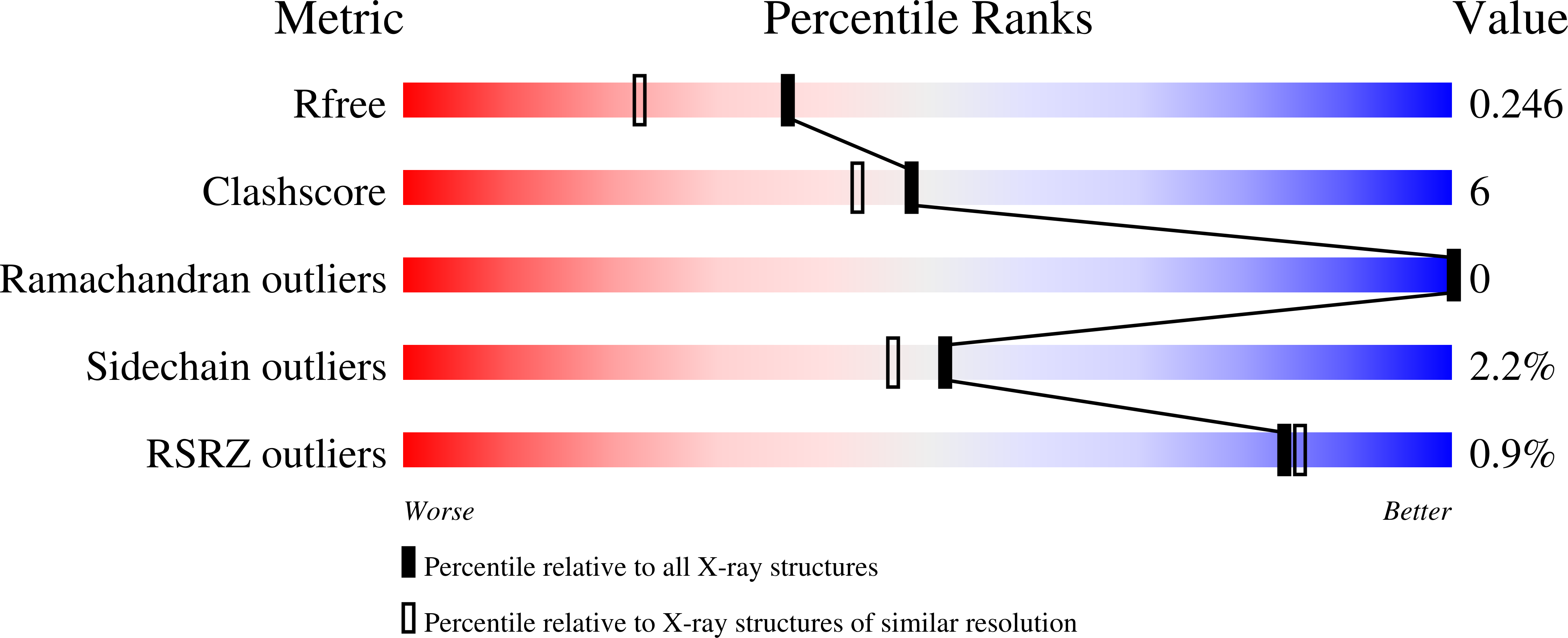
R-Value Free:
0.23
R-Value Work:
0.21
Space Group:
P 1 21 1