
Deposition Date
2022-01-12
Release Date
2022-03-30
Last Version Date
2023-10-18
Entry Detail
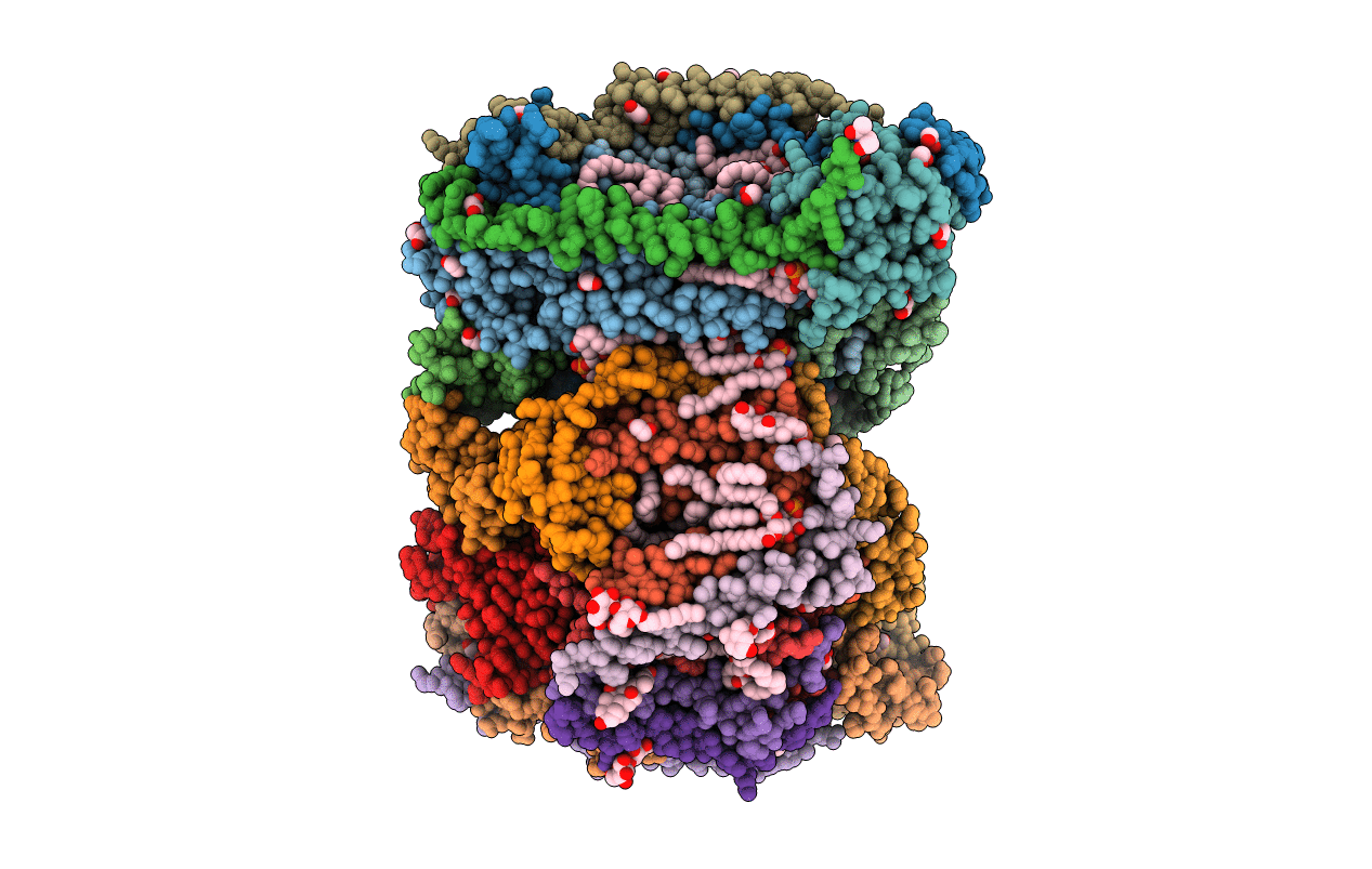
PDB ID:
7THU
Keywords:
Title:
Structure of reduced bovine cytochrome c oxidase at 1.93 Angstrom resolution obtained by synchrotron X-rays
Biological Source:
Source Organism(s):
Bos taurus (Taxon ID: 9913)
Method Details:
Experimental Method:
Resolution:
1.93 Å
R-Value Free:
0.21
R-Value Work:
0.18
Space Group:
P 21 21 21
