Deposition Date
2022-01-11
Release Date
2022-03-02
Last Version Date
2024-11-06
Entry Detail
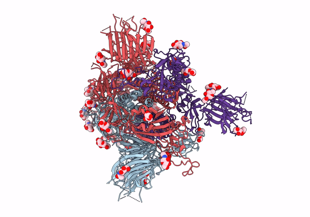
PDB ID:
7THK
Keywords:
Title:
Cryo-EM structure of prefusion SARS-CoV-2 spike omicron B.1.1.529 variant
Biological Source:
Source Organism(s):
Expression System(s):
Method Details:
Experimental Method:
Resolution:
3.11 Å
Aggregation State:
PARTICLE
Reconstruction Method:
SINGLE PARTICLE