Deposition Date
2021-12-22
Release Date
2022-06-22
Last Version Date
2024-11-06
Entry Detail
PDB ID:
7TBP
Keywords:
Title:
X-ray structure of the HIV-1 myristoylated matrix protein
Biological Source:
Source Organism(s):
Human immunodeficiency virus 1 (Taxon ID: 11676)
Expression System(s):
Method Details:
Experimental Method:
Resolution:
2.15 Å
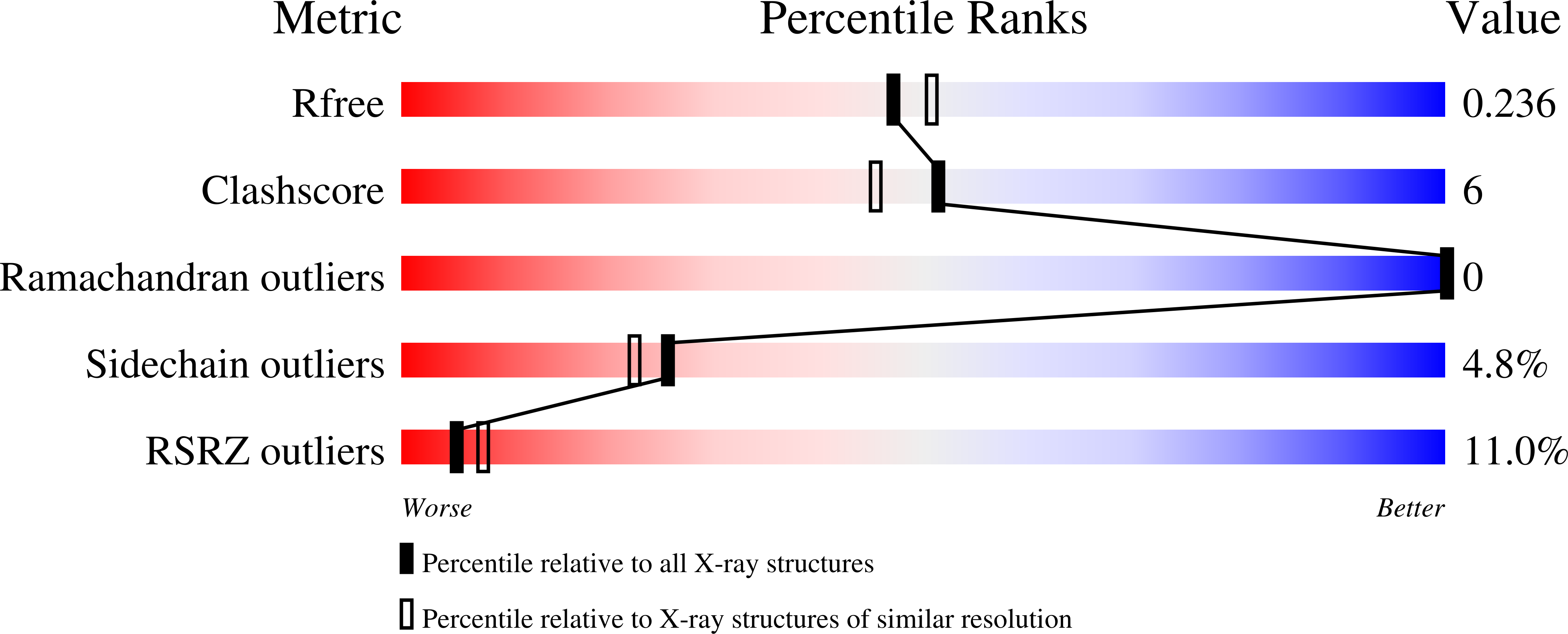
R-Value Free:
0.23
R-Value Work:
0.18
R-Value Observed:
0.18
Space Group:
H 3 2