7T8N image
Entry Detail
PDB ID:
7T8N
Title:
Crystal structure of the PNAG binding module PgaA-TPR 220-359
Biological Source:
Source Organism:
Host Organism:
PDB Version:
Deposition Date:
2021-12-16
Release Date:
2022-08-03
Method Details:
Experimental Method:
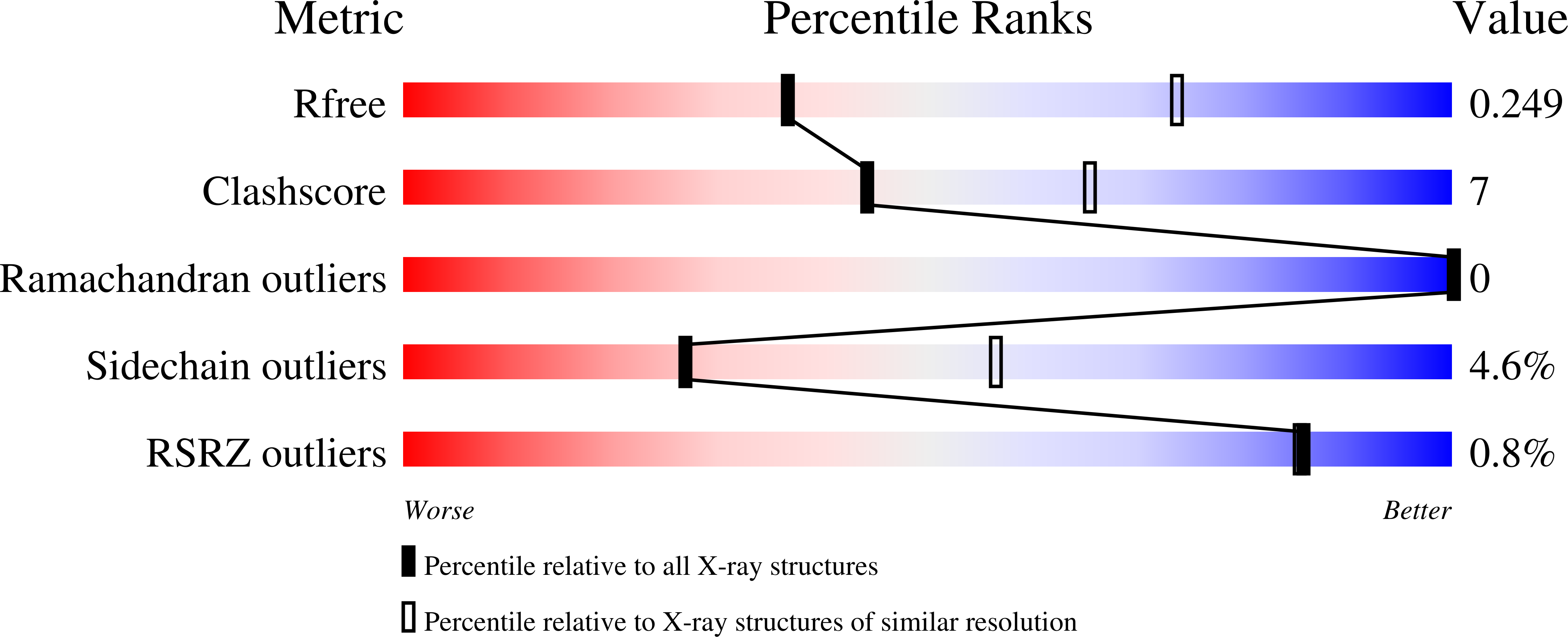
Resolution:
2.85 Å
R-Value Free:
0.26
R-Value Work:
0.23
Space Group:
P 62
Macromolecular Entities
Polymer Type:polypeptide(L)
Description:Poly-beta-1,6-N-acetyl-D-glucosamine export protein
Chain IDs:A (auth: AAA), B (auth: BBB)
Chain Length:148
Number of Molecules:2
Biological Source:Escherichia coli K-12
Primary Citation
The TPR domain of PgaA is a multifunctional scaffold that binds PNAG and modulates PgaB-dependent polymer processing.
Plos Pathog. 18 e1010750 e1010750 (2022)
PMID: 35930610 DOI: 10.1371/journal.ppat.1010750

Abstact

The synthesis of exopolysaccharides as biofilm matrix components by pathogens is a crucial factor for chronic infections and antibiotic resistance. Many periplasmic proteins involved in polymer processing and secretion in Gram-negative synthase dependent exopolysaccharide biosynthetic systems have been individually characterized. The operons responsible for the production of PNAG, alginate, cellulose and the Pel polysaccharide each contain a gene that encodes an outer membrane associated tetratricopeptide repeat (TPR) domain containing protein. While the TPR domain has been shown to bind other periplasmic proteins, the functional consequences of these interactions for the polymer remain poorly understood. Herein, we show that the C-terminal TPR region of PgaA interacts with the de-N-acetylase domain of PgaB, and increases its deacetylase activity. Additionally, we found that when the two proteins form a complex, the glycoside hydrolase activity of PgaB is also increased. To better understand structure-function relationships we determined the crystal structure of a stable TPR module, which has a conserved groove formed by three repeat motifs. Tryptophan quenching, mass spectrometry analysis and molecular dynamics simulation studies suggest that the crystallized TPR module can bind PNAG/dPNAG via its electronegative groove on the concave surface, and potentially guide the polymer through the periplasm towards the porin for export. Our results suggest a scaffolding role for the TPR domain that combines PNAG/dPNAG translocation with the modulation of its chemical structure by PgaB.

Legend

Protein

Chemical

Disease

Primary Citation of related structures