7SV2 image
Deposition Date 2021-11-18
Release Date 2022-05-18
Last Version Date 2023-10-18
Entry Detail
PDB ID:
7SV2
Title:
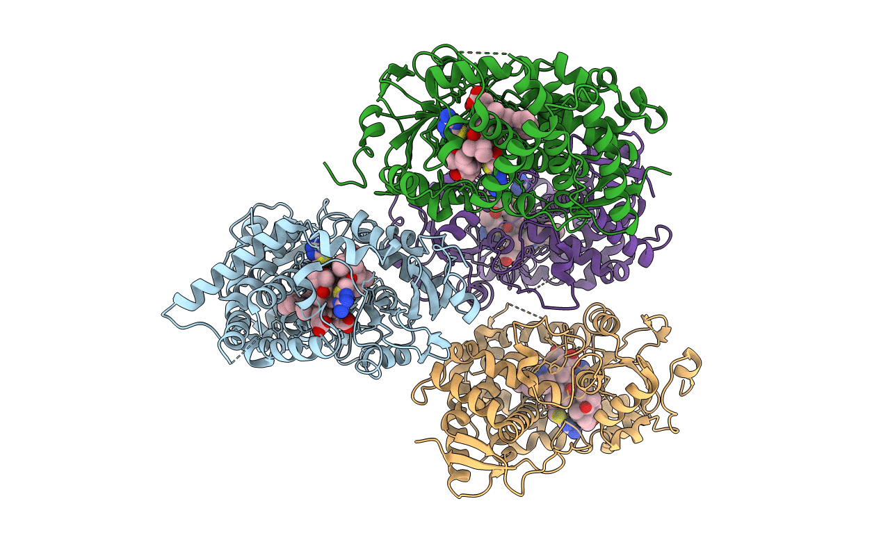
Human Cytochrome P450 (CYP) 3A5 ternary complex with azamulin
Biological Source:
Source Organism:
Homo sapiens (Taxon ID: 9606)
Host Organism:
Method Details:
Experimental Method:
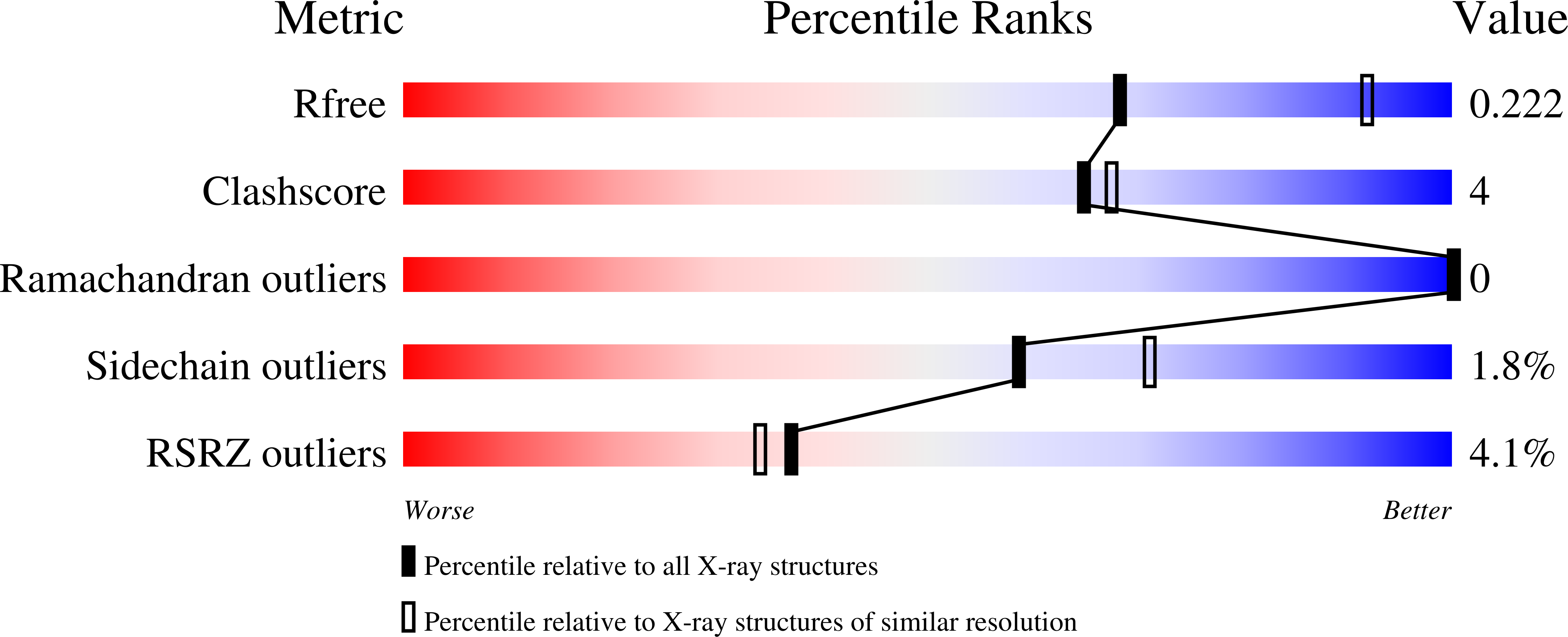
Resolution:
2.46 Å
R-Value Free:
0.22
R-Value Work:
0.18
R-Value Observed:
0.18
Space Group:
C 2 2 21
Macromolecular Entities
Polymer Type:polypeptide(L)
Molecule:Cytochrome P450 3A5
Gene (Uniprot):CYP3A5
Chain IDs:A, B, C, D
Chain Length:480
Number of Molecules:4
Biological Source:Homo sapiens
Primary Citation
Structural characterization of the homotropic cooperative binding of azamulin to human cytochrome P450 3A5.
J.Biol.Chem. 298 101909 101909 (2022)
PMID: 35398097 DOI: 10.1016/j.jbc.2022.101909

Abstact

Cytochrome P450 3A4 and 3A5 catalyze the metabolic clearance of a large portion of therapeutic drugs. Azamulin is used as a selective inhibitor for 3A4 and 3A5 to define their roles in metabolism of new chemical entities during drug development. In contrast to 3A4, 3A5 exhibits homotropic cooperativity for the sequential binding of two azamulin molecules at concentrations used for inhibition. To define the underlying sites and mechanisms for cooperativity, an X-ray crystal structure of 3A5 was determined with two azamulin molecules in the active site that are stacked in an antiparallel orientation. One azamulin resides proximal to the heme in a pose similar to the 3A4-azamulin complex. Comparison to the 3A5 apo structure indicates that the distal azamulin in 3A5 ternary complex causes a significant induced fit that excludes water from the hydrophobic surfaces of binding cavity and the distal azamulin, which is augmented by the stacking interaction with the proximal azamulin. Homotropic cooperativity was not observed for the binding of related pleuromutilin antibiotics, tiamulin, retapamulin, and lefamulin, to 3A5, which are larger and unlikely to bind in the distal site in a stacked orientation. Formation of the 3A5 complex with two azamulin molecules may prevent time-dependent inhibition that is seen for 3A4 by restricting alternate product formation and/or access of reactive intermediates to vulnerable protein sites. These results also contribute to a better understanding of sites for cooperative binding and the differential structural plasticity of 3A5 and 3A4 that contribute to differential substrate and inhibitor binding.

Legend

Protein

Chemical

Disease

Primary Citation of related structures