Deposition Date
2021-11-12
Release Date
2022-05-11
Last Version Date
2024-10-23
Entry Detail
PDB ID:
7ST8
Keywords:
Title:
Crystal structure of 7H2.2 Fab in complex with SAS1B C-terminal region
Biological Source:
Source Organism(s):
Mus musculus (Taxon ID: 10090)
Homo sapiens (Taxon ID: 9606)
Homo sapiens (Taxon ID: 9606)
Expression System(s):
Method Details:
Experimental Method:
Resolution:
2.75 Å
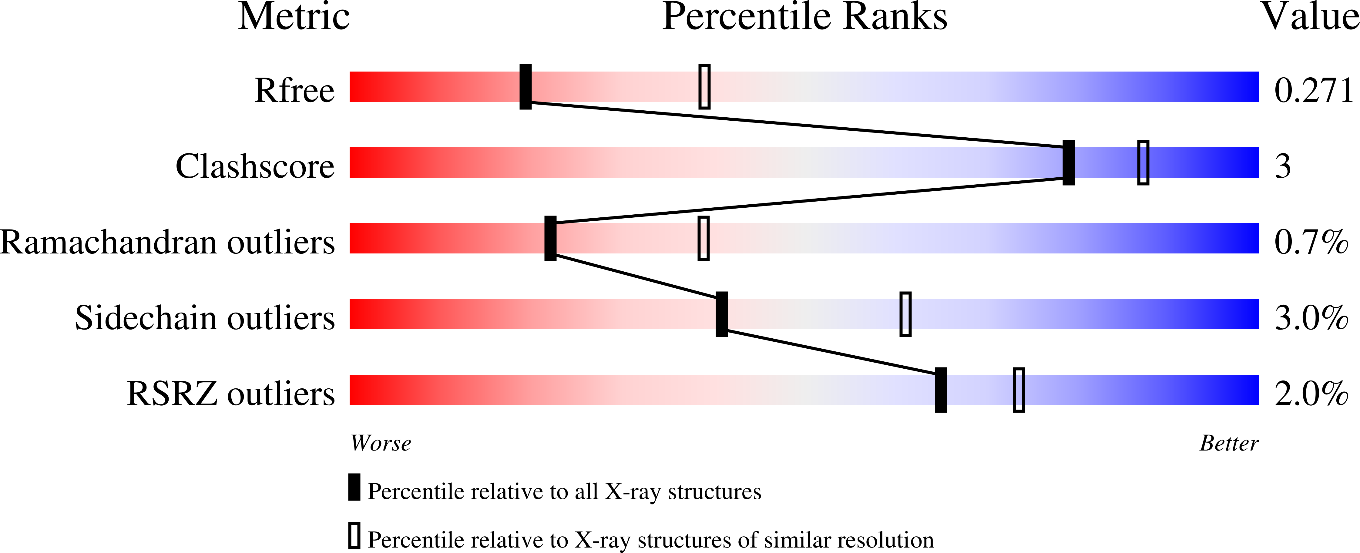
R-Value Free:
0.26
R-Value Work:
0.23
R-Value Observed:
0.23
Space Group:
C 1 2 1