7SPX image
Entry Detail
PDB ID:
7SPX
Title:
Crystal structure of photoactive yellow protein (PYP); F28oCNF construct
Biological Source:
PDB Version:
Deposition Date:
2021-11-03
Release Date:
2022-05-11
Method Details:
Experimental Method:
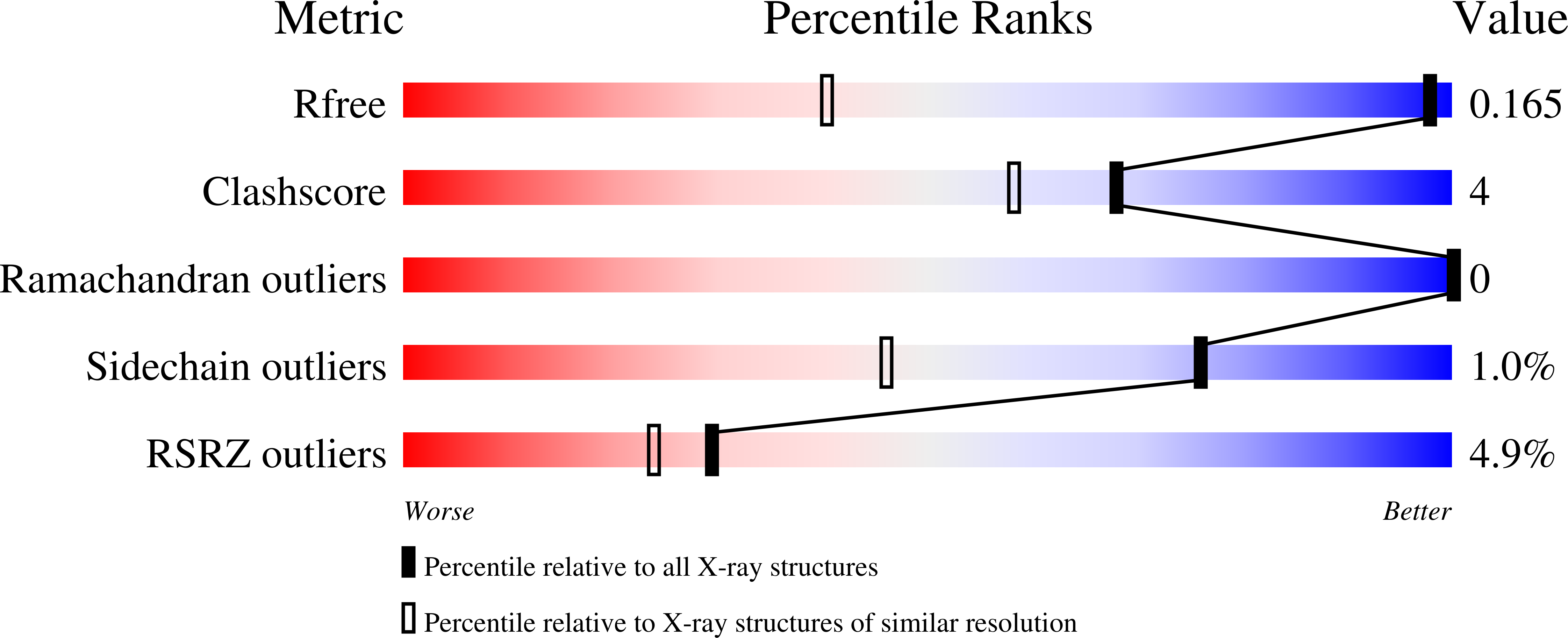
Resolution:
0.97 Å
R-Value Free:
0.16
R-Value Work:
0.14
R-Value Observed:
0.14
Space Group:
P 63
Macromolecular Entities
Polymer Type:polypeptide(L)
Description:Photoactive yellow protein
Chain IDs:A
Chain Length:125
Number of Molecules:1
Biological Source:Halorhodospira halophila
Modified Residue
Compound ID Chain ID Parent Comp ID Details 2D Image
9IJ A PHE modified residue
Ligand Molecules
Primary Citation
Nitrile Infrared Intensities Characterize Electric Fields and Hydrogen Bonding in Protic, Aprotic, and Protein Environments.
J.Am.Chem.Soc. 144 7562 7567 (2022)
PMID: 35467853 DOI: 10.1021/jacs.2c00675

Abstact

Nitriles are widely used vibrational probes; however, the interpretation of their IR frequencies is complicated by hydrogen bonding (H-bonding) in protic environments. We report a new vibrational Stark effect (VSE) that correlates the electric field projected on the -C≡N bond to the transition dipole moment and, by extension, the nitrile peak area or integrated intensity. This linear VSE applies to both H-bonding and non-H-bonding interactions. It can therefore be generally applied to determine electric fields in all environments. Additionally, it allows for semiempirical extraction of the H-bonding contribution to the blueshift of the nitrile frequency. Nitriles were incorporated at H-bonding and non-H-bonding protein sites using amber suppression, and each nitrile variant was structurally characterized at high resolution. We exploited the combined information available from variations in frequency and integrated intensity and demonstrate that nitriles are a generally useful probe for electric fields.

Legend

Protein

Chemical

Disease

Primary Citation of related structures