7SNB image
Deposition Date 2021-10-27
Release Date 2022-03-02
Last Version Date 2025-02-26
Entry Detail
PDB ID:
7SNB
Keywords:
Title:
The X-ray crystal structure of the N-terminal domain of Staphylococcus aureus Fatty Acid Kinase A (FakA, residues 1-208) in complex with AMP and ADP to 1.105 Angstrom resolution
Biological Source:
Source Organism:
Host Organism:
Method Details:
Experimental Method:
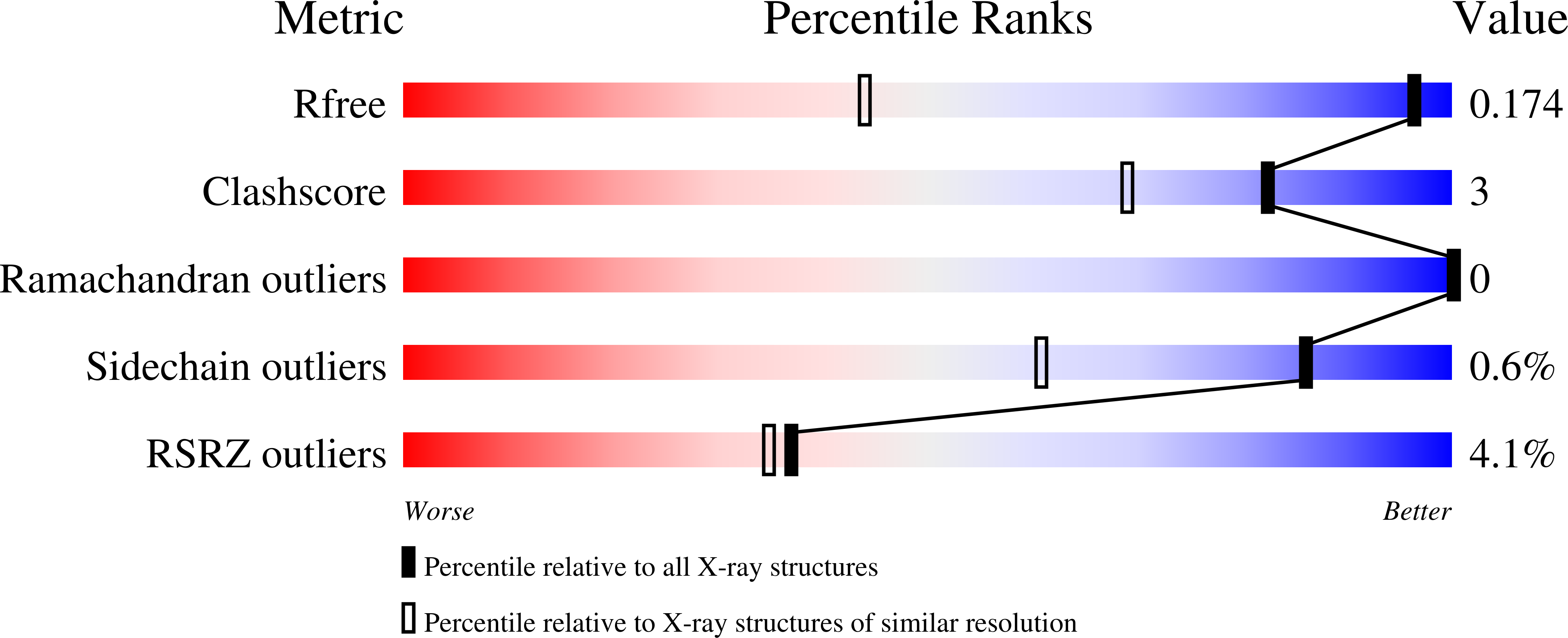
Resolution:
1.11 Å
R-Value Free:
0.17
R-Value Work:
0.15
R-Value Observed:
0.15
Space Group:
P 21 21 21
Macromolecular Entities
Polymer Type:polypeptide(L)
Molecule:Fatty Acid Kinase A
Chain IDs:A
Chain Length:228
Number of Molecules:1
Biological Source:Staphylococcus aureus
Primary Citation
Domain architecture and catalysis of the Staphylococcus aureus fatty acid kinase.
J.Biol.Chem. 298 101993 101993 (2022)
PMID: 35490779 DOI: 10.1016/j.jbc.2022.101993

Abstact

Fatty acid kinase (Fak) is a two-component enzyme that generates acyl-phosphate for phospholipid synthesis. Fak consists of a kinase domain protein (FakA) that phosphorylates a fatty acid enveloped by a fatty acid binding protein (FakB). The structural basis for FakB function has been established, but little is known about FakA. Here, we used limited proteolysis to define three separate FakA domains: the amino terminal FakA_N, the central FakA_L, and the carboxy terminal FakA_C. The isolated domains lack kinase activity, but activity is restored when FakA_N and FakA_L are present individually or connected as FakA_NL. The X-ray structure of the monomeric FakA_N captures the product complex with ADP and two Mg2+ ions bound at the nucleotide site. The FakA_L domain encodes the dimerization interface along with conserved catalytic residues Cys240, His282, and His284. AlphaFold analysis of FakA_L predicts the catalytic residues are spatially clustered and pointing away from the dimerization surface. Furthermore, the X-ray structure of FakA_C shows that it consists of two subdomains that are structurally related to FakB. Analytical ultracentrifugation demonstrates that FakA_C binds FakB, and site-directed mutagenesis confirms that a positively charged wedge on FakB meshes with a negatively charged groove on FakA_C. Finally, small angle X-ray scattering analysis is consistent with freely rotating FakA_N and FakA_C domains tethered by flexible linkers to FakA_L. These data reveal specific roles for the three independently folded FakA protein domains in substrate binding and catalysis.

Legend

Protein

Chemical

Disease

Primary Citation of related structures