7SKT image
Entry Detail
PDB ID:
7SKT
Keywords:
Title:
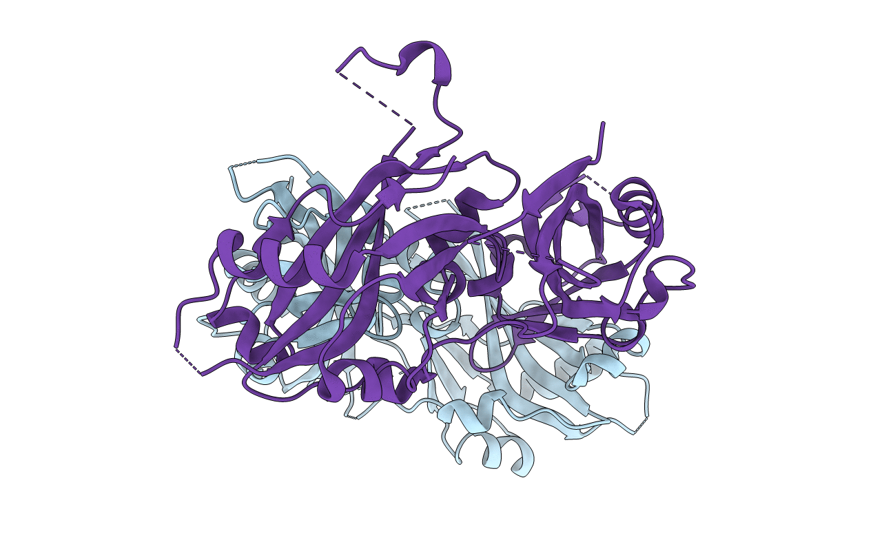
Crystal structure of Nipah virus matrix protein
Biological Source:
Source Organism:
Host Organism:
PDB Version:
Deposition Date:
2021-10-21
Release Date:
2022-08-24
Method Details:
Experimental Method:
Resolution:
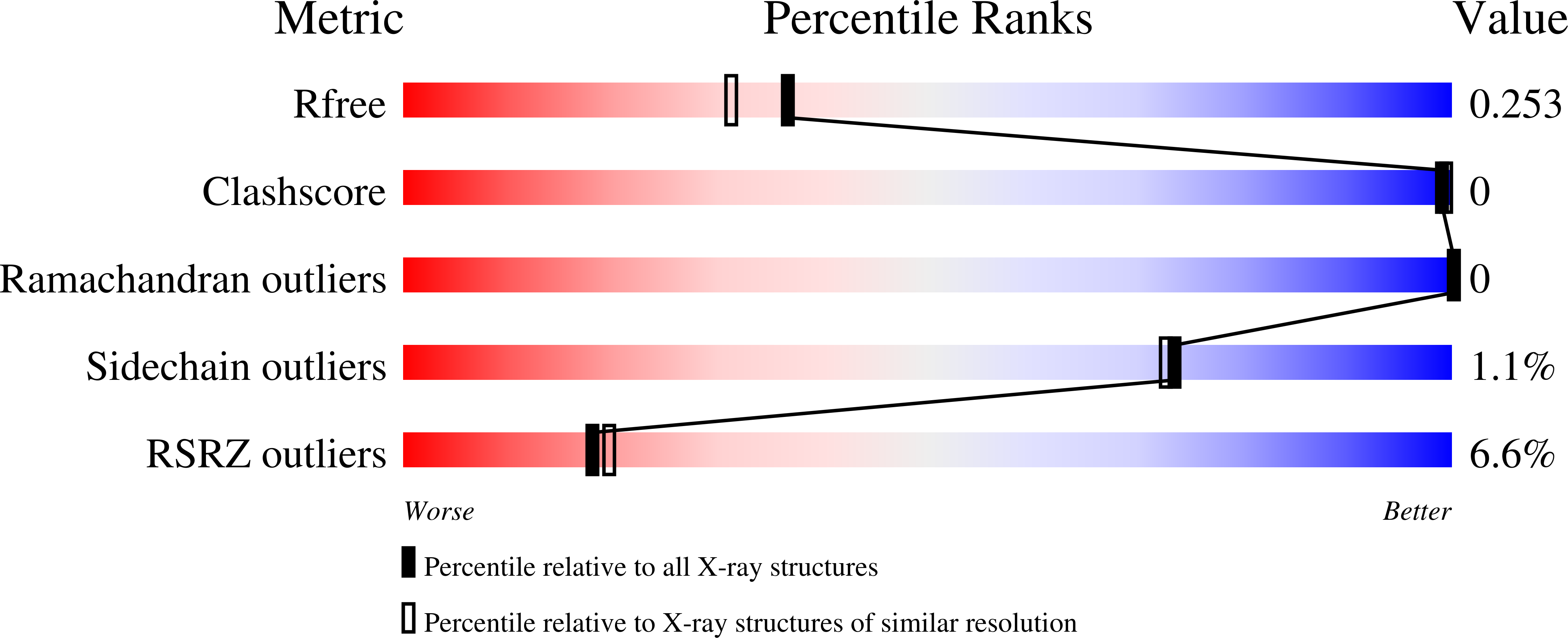
2.05 Å
R-Value Free:
0.25
R-Value Work:
0.21
R-Value Observed:
0.21
Space Group:
P 21 21 21
Macromolecular Entities
Polymer Type:polypeptide(L)
Description:Matrix protein
Chain IDs:A, B
Chain Length:393
Number of Molecules:2
Biological Source:Nipah henipavirus
Primary Citation
Measles and Nipah virus assembly: Specific lipid binding drives matrix polymerization.
Sci Adv 8 eabn1440 eabn1440 (2022)
PMID: 35857835 DOI: 10.1126/sciadv.abn1440

Abstact

Measles virus, Nipah virus, and multiple other paramyxoviruses cause disease outbreaks in humans and animals worldwide. The paramyxovirus matrix (M) protein mediates virion assembly and budding from host cell membranes. M is thus a key target for antivirals, but few high-resolution structures of paramyxovirus M are available, and we lack the clear understanding of how viral M proteins interact with membrane lipids to mediate viral assembly and egress that is needed to guide antiviral design. Here, we reveal that M proteins associate with phosphatidylserine and phosphatidylinositol 4,5-bisphosphate [PI(4,5)P2] at the plasma membrane. Using x-ray crystallography, electron microscopy, and molecular dynamics, we demonstrate that PI(4,5)P2 binding induces conformational and electrostatic changes in the M protein surface that trigger membrane deformation, matrix layer polymerization, and virion assembly.

Legend

Protein

Chemical

Disease

Primary Citation of related structures