Deposition Date
2021-10-04
Release Date
2021-10-27
Last Version Date
2023-10-18
Entry Detail
PDB ID:
7SG3
Keywords:
Title:
The X-ray crystal structure of the Staphylococcus aureus Fatty Acid Kinase B1 mutant A121I-A158L to 2.35 Angstrom resolution (Open form chain A, Palmitate bound)
Biological Source:
Source Organism(s):
Staphylococcus aureus (Taxon ID: 1280)
Expression System(s):
Method Details:
Experimental Method:
Resolution:
2.35 Å
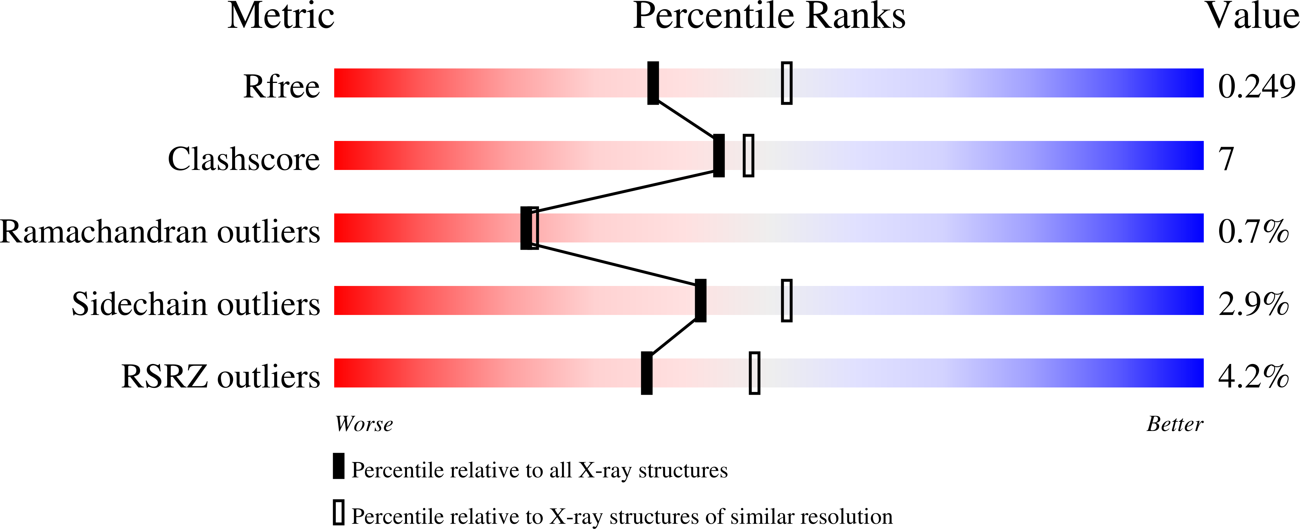
R-Value Free:
0.25
R-Value Work:
0.21
R-Value Observed:
0.21
Space Group:
P 1