Deposition Date
2021-10-03
Release Date
2022-04-27
Last Version Date
2024-10-09
Entry Detail
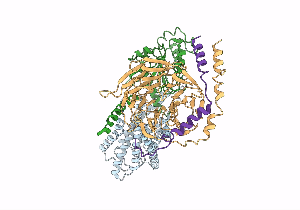
PDB ID:
7SF7
Keywords:
Title:
LPHN3 (ADGRL3) 7TM domain bound to tethered agonist in complex with G protein heterotrimer
Biological Source:
Source Organism(s):
Homo sapiens (Taxon ID: 9606)
Expression System(s):
Method Details:
Experimental Method:
Resolution:
2.90 Å
Aggregation State:
PARTICLE
Reconstruction Method:
SINGLE PARTICLE