7SF0 image
Entry Detail
PDB ID:
7SF0
Keywords:
Title:
Crystal structure of Vaccinia Virus decapping enzyme D9 in complex with trinucleotide substrate
Biological Source:
PDB Version:
Deposition Date:
2021-10-02
Release Date:
2022-03-30
Method Details:
Experimental Method:
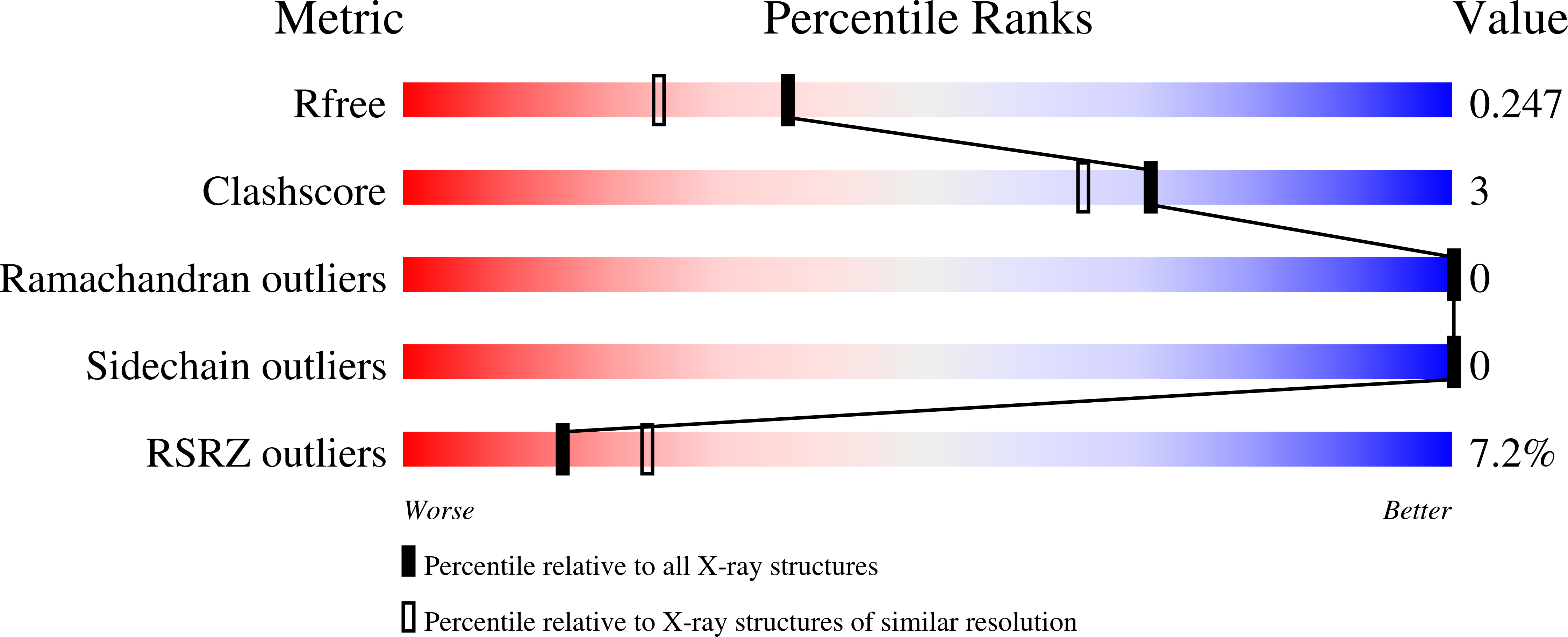
Resolution:
1.95 Å
R-Value Free:
0.24
R-Value Work:
0.20
R-Value Observed:
0.20
Space Group:
C 2 2 21
Macromolecular Entities
Polymer Type:polypeptide(L)
Description:DNA repair NTP-phosphohydrolase
Chain IDs:A
Chain Length:221
Number of Molecules:1
Biological Source:Vaccinia virus Western Reserve
Primary Citation
Structure of the poxvirus decapping enzyme D9 reveals its mechanism of cap recognition and catalysis.
Structure 30 721 ? (2022)
PMID: 35290794 DOI: 10.1016/j.str.2022.02.012

Abstact

Poxviruses encode decapping enzymes that remove the protective 5' cap from both host and viral mRNAs to commit transcripts for decay by the cellular exonuclease Xrn1. Decapping by these enzymes is critical for poxvirus pathogenicity by means of simultaneously suppressing host protein synthesis and limiting the accumulation of viral double-stranded RNA (dsRNA), a trigger for antiviral responses. Here we present a high-resolution structural view of the vaccinia virus decapping enzyme D9. This Nudix enzyme contains a domain organization different from other decapping enzymes in which a three-helix bundle is inserted into the catalytic Nudix domain. The 5' mRNA cap is positioned in a bipartite active site at the interface of the two domains. Specificity for the methylated guanosine cap is achieved by stacking between conserved aromatic residues in a manner similar to that observed in canonical cap-binding proteins VP39, eIF4E, and CBP20, and distinct from eukaryotic decapping enzyme Dcp2.

Legend

Protein

Chemical

Disease

Primary Citation of related structures