7SD3 image
Deposition Date 2021-09-29
Release Date 2022-11-09
Last Version Date 2024-11-06
Entry Detail
PDB ID:
7SD3
Title:
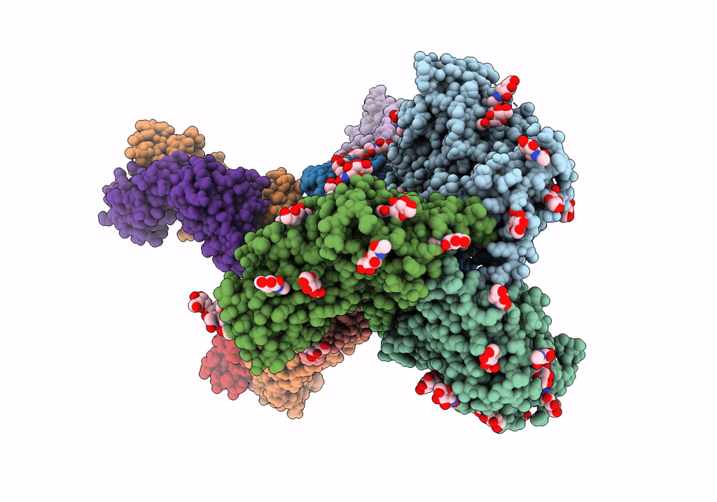
Cytoplasmic tail deleted HIV-1 Env bound with three 4E10 Fabs
Biological Source:
Source Organism:
Host Organism:
Method Details:
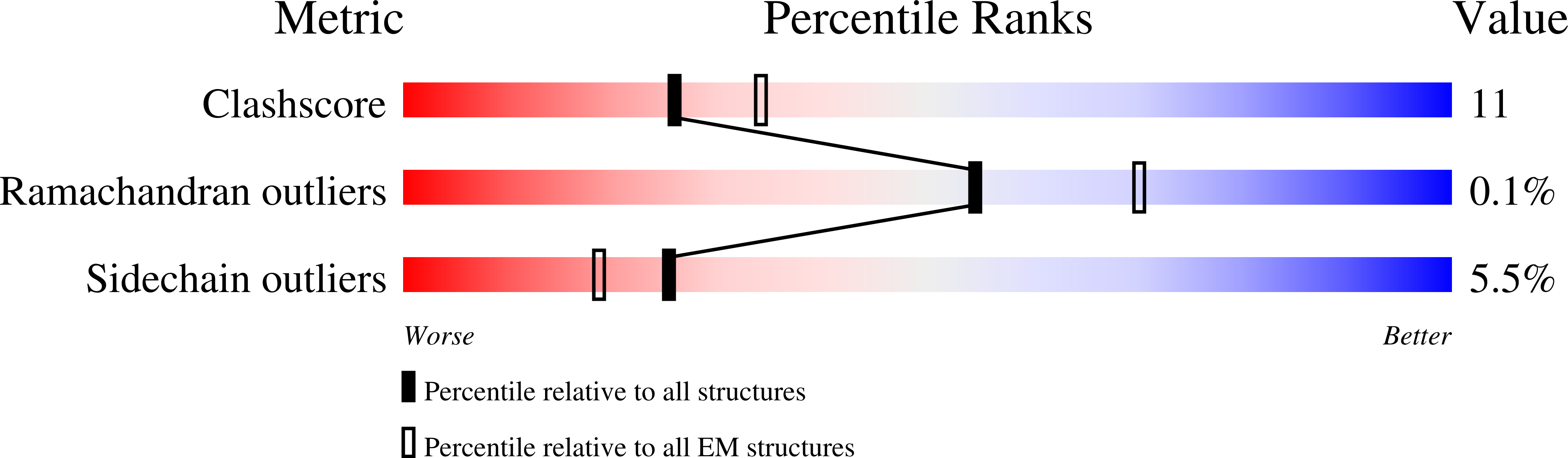
Experimental Method:
Resolution:
3.67 Å
Aggregation State:
PARTICLE
Reconstruction Method:
SINGLE PARTICLE
Macromolecular Entities
Polymer Type:polypeptide(L)
Molecule:Envelope glycoprotein gp120
Chain IDs:G (auth: A), I (auth: C), K (auth: E)
Chain Length:481
Number of Molecules:3
Biological Source:HIV whole-genome vector AA1305#18
Polymer Type:polypeptide(L)
Molecule:Transmembrane protein gp41
Chain IDs:H (auth: B), J (auth: D), L (auth: F)
Chain Length:173
Number of Molecules:3
Biological Source:HIV whole-genome vector AA1305#18
Polymer Type:polypeptide(L)
Molecule:4E10 Fab heavy chain
Chain IDs:B (auth: G), D (auth: K), F (auth: I)
Chain Length:230
Number of Molecules:3
Biological Source:Homo sapiens
Polymer Type:polypeptide(L)
Molecule:4E10 Fab light chain
Chain IDs:A (auth: H), C (auth: N), E (auth: M)
Chain Length:214
Number of Molecules:3
Biological Source:Homo sapiens
Ligand Molecules
Primary Citation
Dynamic HIV-1 spike motion creates vulnerability for its membrane-bound tripod to antibody attack.
Nat Commun 13 6393 6393 (2022)
PMID: 36302771 DOI: 10.1038/s41467-022-34008-y

Abstact

Vaccines targeting HIV-1's gp160 spike protein are stymied by high viral mutation rates and structural chicanery. gp160's membrane-proximal external region (MPER) is the target of naturally arising broadly neutralizing antibodies (bnAbs), yet MPER-based vaccines fail to generate bnAbs. Here, nanodisc-embedded spike protein was investigated by cryo-electron microscopy and molecular-dynamics simulations, revealing spontaneous ectodomain tilting that creates vulnerability for HIV-1. While each MPER protomer radiates centrally towards the three-fold axis contributing to a membrane-associated tripod structure that is occluded in the upright spike, tilting provides access to the opposing MPER. Structures of spike proteins with bound 4E10 bnAb Fabs reveal that the antibody binds exposed MPER, thereby altering MPER dynamics, modifying average ectodomain tilt, and imposing strain on the viral membrane and the spike's transmembrane segments, resulting in the abrogation of membrane fusion and informing future vaccine development.

Legend

Protein

Chemical

Disease

Primary Citation of related structures