7SCX image
Entry Detail
PDB ID:
7SCX
Keywords:
Title:
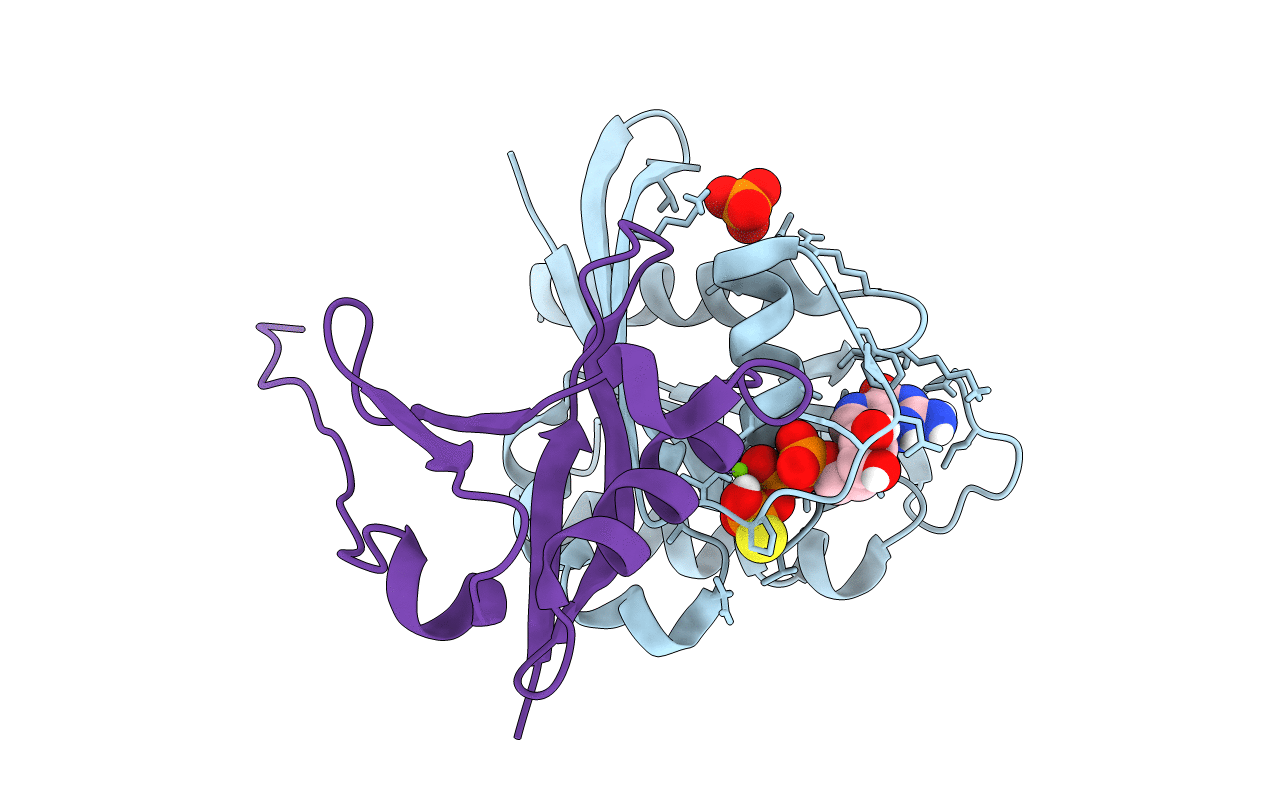
KRAS full-length G12V in complex with RGL1 Ras association domain
Biological Source:
Source Organism:
Host Organism:
PDB Version:
Deposition Date:
2021-09-29
Release Date:
2022-05-18
Method Details:
Experimental Method:
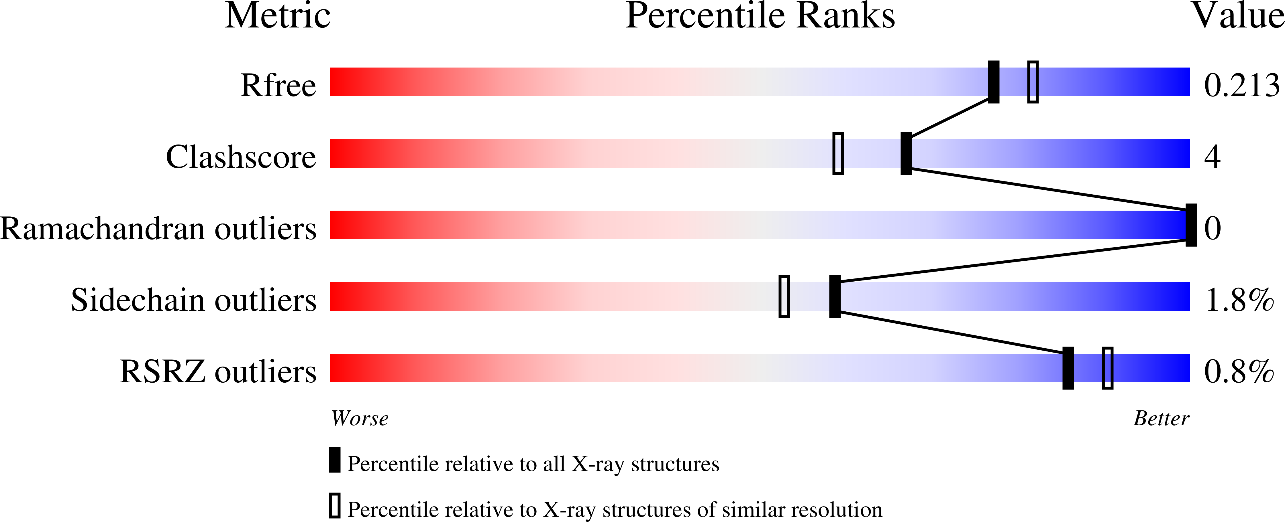
Resolution:
1.96 Å
R-Value Free:
0.21
R-Value Work:
0.17
R-Value Observed:
0.17
Space Group:
C 1 2 1
Macromolecular Entities
Polymer Type:polypeptide(L)
Description:Isoform 2B of GTPase KRas
Mutations:G12V
Chain IDs:A
Chain Length:188
Number of Molecules:1
Biological Source:Homo sapiens
Polymer Type:polypeptide(L)
Description:Ral guanine nucleotide dissociation stimulator-like 1
Chain IDs:B
Chain Length:93
Number of Molecules:1
Biological Source:Homo sapiens
Primary Citation
Structures of RGL1 RAS-Association Domain in Complex with KRAS and the Oncogenic G12V Mutant.
J.Mol.Biol. 434 167527 167527 (2022)
PMID: 35257782 DOI: 10.1016/j.jmb.2022.167527

Abstact

Ral Guanine Nucleotide Dissociation Stimulator Like 1 (RGL1) is a RAS effector protein that activates Ral GTPase by stimulating nucleotide exchange. Most structures of RAS-effector complexes are for the HRAS isoform; relatively few KRAS-effector structures have been solved, even though KRAS mutations are more frequent in human cancers. We determined crystal structures of KRAS/RGL1-RAS-association (RA) domain complexes and characterized the interaction in solution using nuclear magnetic resonance spectroscopy, size-exclusion chromatography combined with multi-angle light scattering and biolayer interferometry. We report structures of wild-type KRAS and the oncogenic G12V mutant in complex with the RA domain of RGL1 at < 2 Å resolution. KRASWT/RGL1-RA crystallized as a 1:1 heterodimer, whilst KRASG12V/RGL1-RA crystallized as a heterotetrameric structure in which RGL1-RA dimerized via domain-swapping the C-terminal beta-strand. Solution data indicated that KRASWT and KRASG12V in complex with RGL1-RA both exist predominantly as 1:1 dimers, while tetramerization occurs through very slow association. Through detailed structural analyses, the distance and angle between RAS α1 helix and RBD/RA α1 helix were found to differ significantly among RAS and RBD/RA complexes. The KRAS/RGL1-RA structures possess some of the largest α1RAS/α1Effector distances (21.7-22.2 Å), whereas the corresponding distances in previously reported RAS/RAF complexes are significantly shorter (15.2-17.7 Å). Contact map analysis identified unique structural signatures involving contacts between the β1-β2 loop of RA and the α1 helix of RAS, clearly distinguishing the KRAS/RGL1-RA (and other RAS/RA complexes) from RAS/RBD complexes. These results demonstrate that RAS effectors employ an assortment of finely-tuned docking surfaces to achieve optimal interactions with RAS.

Legend

Protein

Chemical

Disease

Primary Citation of related structures