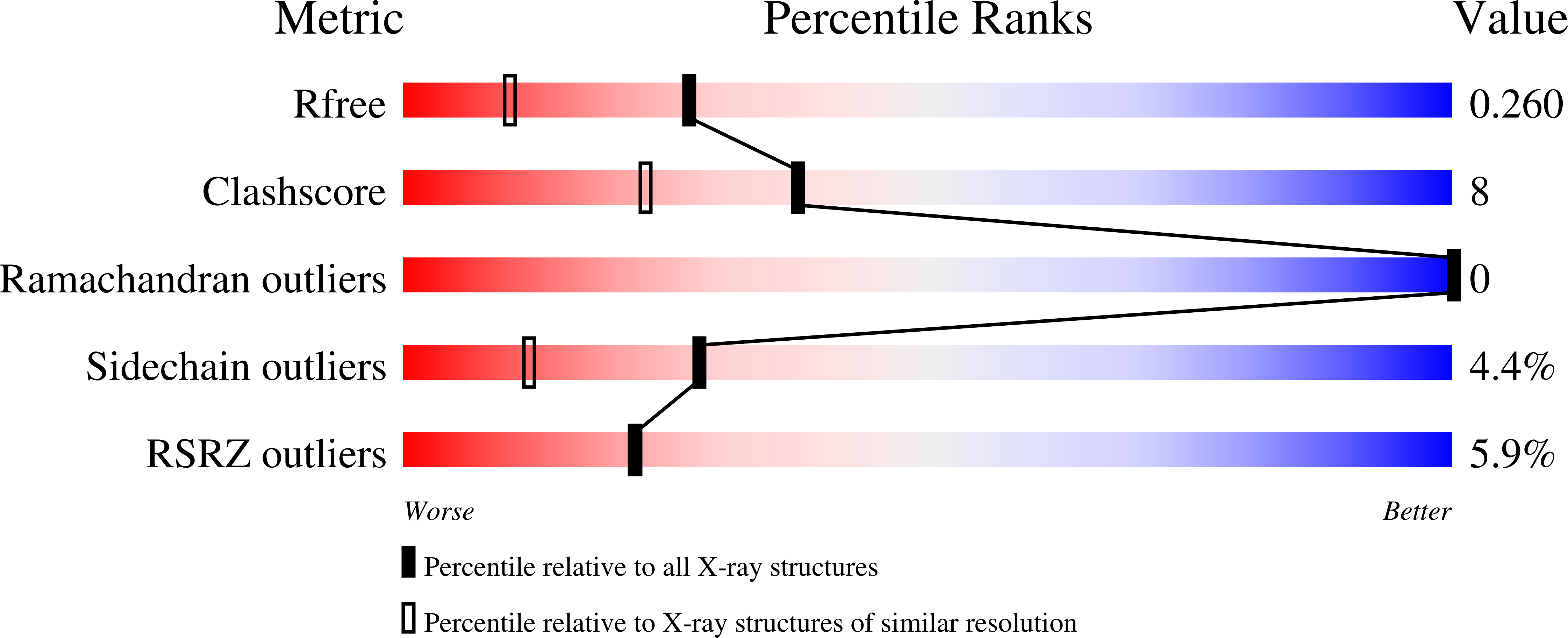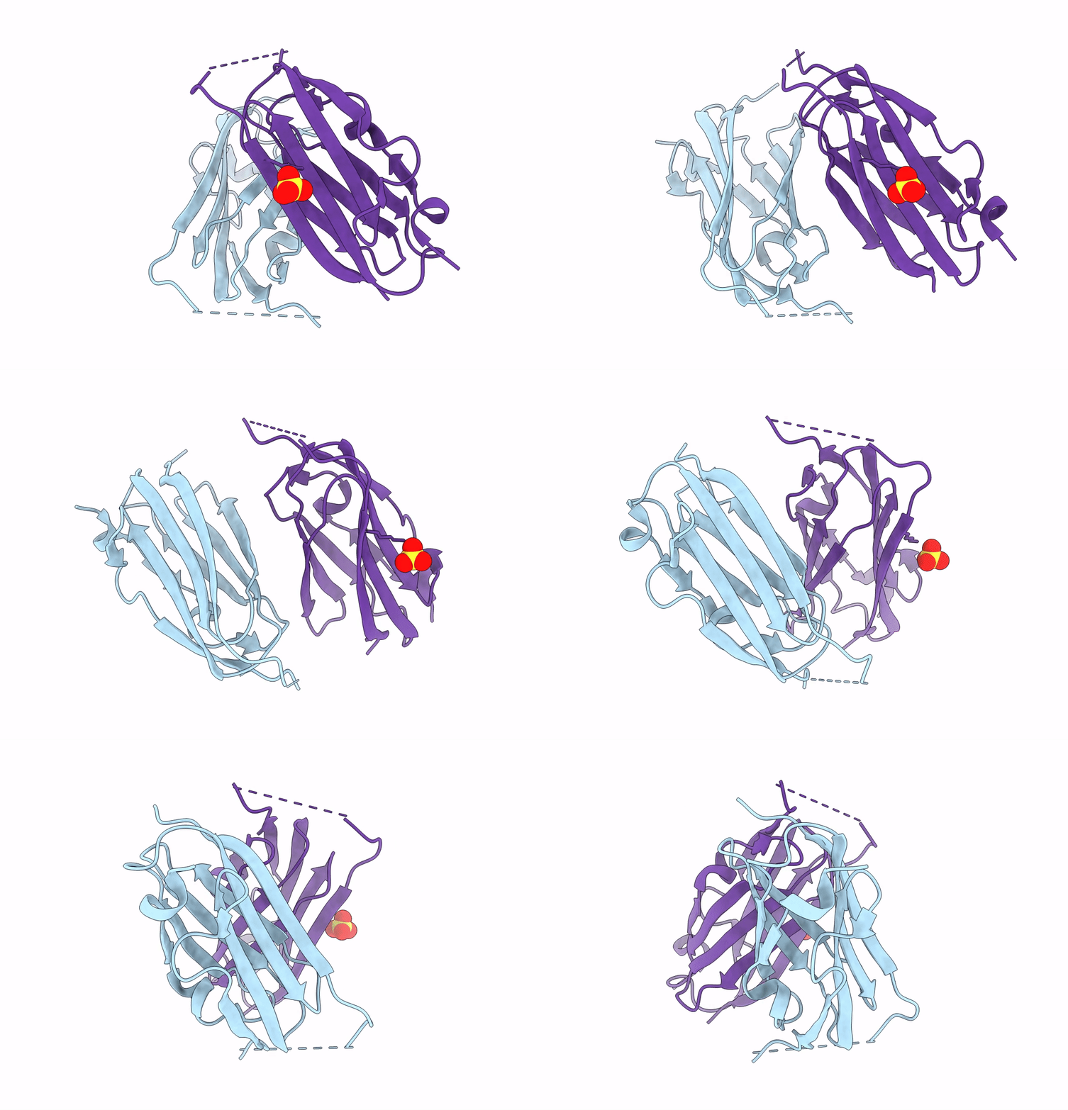
7SC4 image
Entry Detail
PDB ID:
7SC4
Keywords:
Title:
filamin complex-1
Biological Source:
Source Organism:
PDB Version:
Deposition Date:
2021-09-27
Release Date:
2023-04-12
Method Details:
Experimental Method:
Resolution:
1.85 Å
R-Value Free:
0.26
R-Value Work:
0.19
R-Value Observed:
0.20
Space Group:
P 21 2 21
Macromolecular Entities
Polymer Type:polypeptide(L)
Description:Filamin-A/Integrin alpha-IIb light chain, form 2 chimera
Chain IDs:A, B
Chain Length:130
Number of Molecules:2
Biological Source:Homo sapiens
Ligand Molecules
Primary Citation
A mechanism of platelet integrin alpha IIb beta 3 outside-in signaling through a novel integrin alpha IIb subunit-filamin-actin linkage.
Blood 141 2629 2641 (2023)
PMID: 36867840 DOI: 10.1182/blood.2022018333

Abstact

The communication of talin-activated integrin αIIbβ3 with the cytoskeleton (integrin outside-in signaling) is essential for platelet aggregation, wound healing, and hemostasis. Filamin, a large actin crosslinker and integrin binding partner critical for cell spreading and migration, is implicated as a key regulator of integrin outside-in signaling. However, the current dogma is that filamin, which stabilizes inactive αIIbβ3, is displaced from αIIbβ3 by talin to promote the integrin activation (inside-out signaling), and how filamin further functions remains unresolved. Here, we show that while associating with the inactive αIIbβ3, filamin also associates with the talin-bound active αIIbβ3 to mediate platelet spreading. Fluorescence resonance energy transfer-based analysis reveals that while associating with both αIIb and β3 cytoplasmic tails (CTs) to maintain the inactive αIIbβ3, filamin is spatiotemporally rearranged to associate with αIIb CT alone on activated αIIbβ3. Consistently, confocal cell imaging indicates that integrin α CT-linked filamin gradually delocalizes from the β CT-linked focal adhesion marker-vinculin likely because of the separation of integrin α/β CTs occurring during integrin activation. High-resolution crystal and nuclear magnetic resonance structure determinations unravel that the activated integrin αIIb CT binds to filamin via a striking α-helix→β-strand transition with a strengthened affinity that is dependent on the integrin-activating membrane environment containing enriched phosphatidylinositol 4,5-bisphosphate. These data suggest a novel integrin αIIb CT-filamin-actin linkage that promotes integrin outside-in signaling. Consistently, disruption of such linkage impairs the activation state of αIIbβ3, phosphorylation of focal adhesion kinase/proto-oncogene tyrosine kinase Src, and cell migration. Together, our findings advance the fundamental understanding of integrin outside-in signaling with broad implications in blood physiology and pathology.

Legend

Protein

Chemical

Disease

Primary Citation of related structures