
Deposition Date
2021-09-27
Release Date
2021-12-01
Last Version Date
2024-05-22
Entry Detail
PDB ID:
7SC3
Keywords:
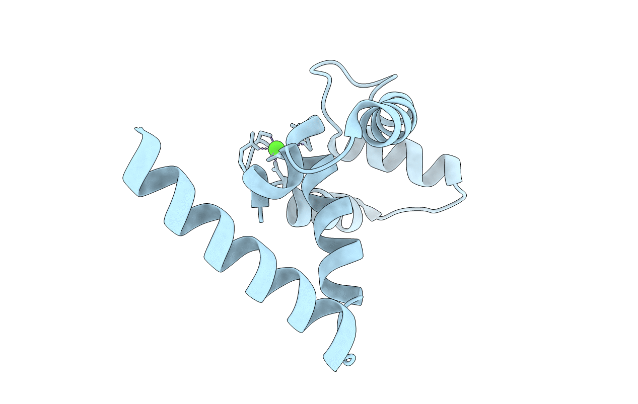
Title:
CRYSTAL STRUCTURE OF THE N-DOMAIN OF CARDIAC MUSCLE TROPONIN C TETHERED TO THE SWITCH REGION OF CARDIAC MUSCLE TROPONIN I (ORTHORHOMBIC FORM)
Biological Source:
Source Organism(s):
Homo sapiens (Taxon ID: 9606)
Expression System(s):
Method Details:
Experimental Method:
Resolution:
2.23 Å
R-Value Free:
0.27
R-Value Work:
0.24
R-Value Observed:
0.24
Space Group:
P 21 21 21
