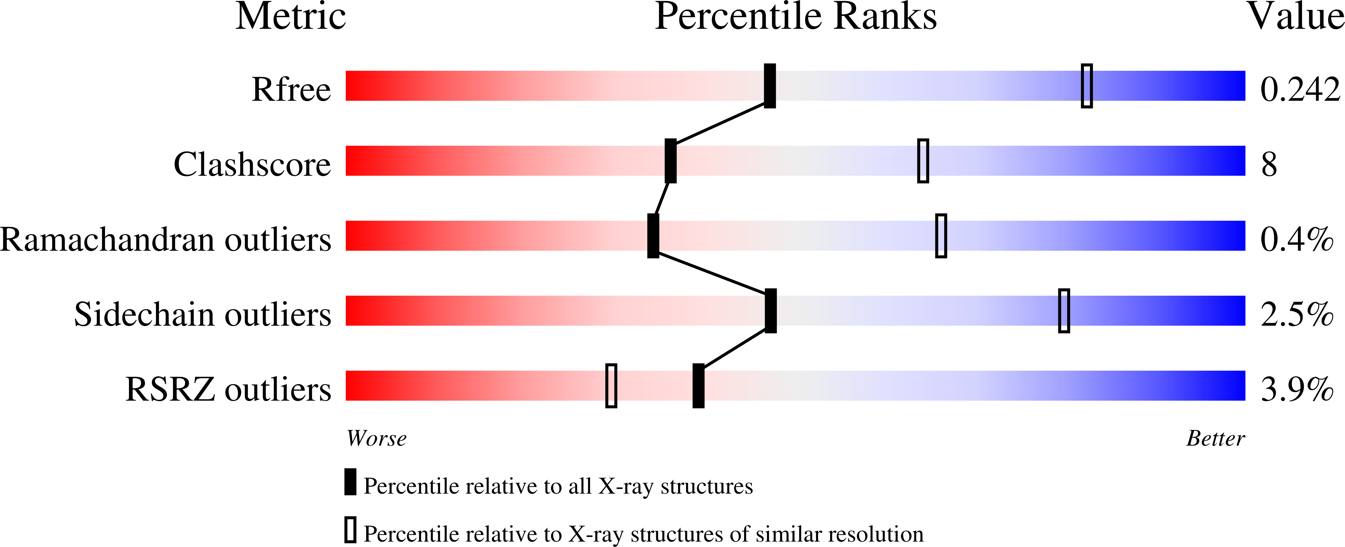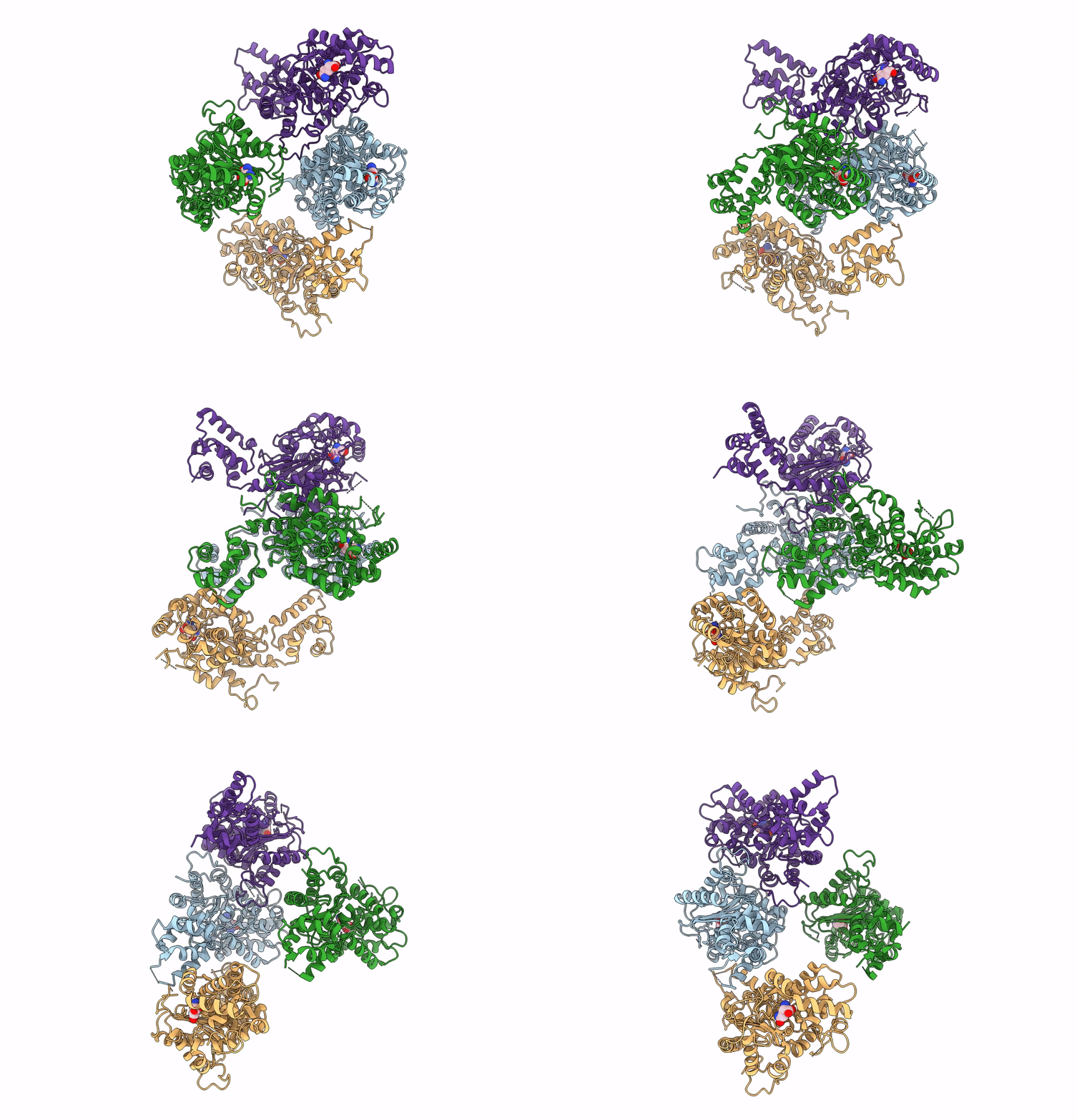
7SBM image
Deposition Date 2021-09-25
Release Date 2022-02-02
Last Version Date 2023-10-18
Entry Detail
PDB ID:
7SBM
Keywords:
Title:
Human glutaminase C (Y466W) with L-Gln, open conformation
Biological Source:
Source Organism:
Homo sapiens (Taxon ID: 9606)
Host Organism:
Method Details:
Experimental Method:
Resolution:
2.80 Å
R-Value Free:
0.24
R-Value Work:
0.20
R-Value Observed:
0.20
Space Group:
P 21 21 21
Macromolecular Entities
Polymer Type:polypeptide(L)
Molecule:Isoform 3 of Glutaminase kidney isoform, mitochondrial
Gene (Uniprot):GLS
Mutations:Y466W
Chain IDs:A, B, C, D
Chain Length:539
Number of Molecules:4
Biological Source:Homo sapiens
Ligand Molecules
Primary Citation
High-resolution structures of mitochondrial glutaminase C tetramers indicate conformational changes upon phosphate binding.
J.Biol.Chem. 298 101564 101564 (2022)
PMID: 34999118 DOI: 10.1016/j.jbc.2022.101564

Abstact

The mitochondrial enzyme glutaminase C (GAC) is upregulated in many cancer cells to catalyze the first step in glutamine metabolism, the hydrolysis of glutamine to glutamate. The dependence of cancer cells on this transformed metabolic pathway highlights GAC as a potentially important therapeutic target. GAC acquires maximal catalytic activity upon binding to anionic activators such as inorganic phosphate. To delineate the mechanism of GAC activation, we used the tryptophan substitution of tyrosine 466 in the catalytic site of the enzyme as a fluorescent reporter for glutamine binding in the presence and absence of phosphate. We show that in the absence of phosphate, glutamine binding to the Y466W GAC tetramer exhibits positive cooperativity. A high-resolution X-ray structure of tetrameric Y466W GAC bound to glutamine suggests that cooperativity in substrate binding is coupled to tyrosine 249, located at the edge of the catalytic site (i.e., the "lid"), adopting two distinct conformations. In one dimer within the GAC tetramer, the lids are open and glutamine binds weakly, whereas, in the adjoining dimer, the lids are closed over the substrates, resulting in higher affinity interactions. When crystallized in the presence of glutamine and phosphate, all four subunits of the Y466W GAC tetramer exhibited bound glutamine with closed lids. Glutamine can bind with high affinity to each subunit, which subsequently undergo simultaneous catalysis. These findings explain how the regulated transitioning of GAC between different conformational states ensures that maximal catalytic activity is reached in cancer cells only when an allosteric activator is available.

Legend

Protein

Chemical

Disease

Primary Citation of related structures