Deposition Date
2021-09-14
Release Date
2021-12-29
Last Version Date
2023-10-18
Entry Detail
PDB ID:
7S6S
Keywords:
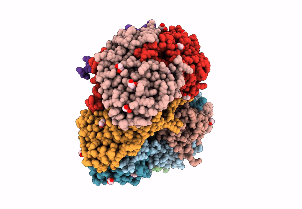
Title:
Complex structure of Methane monooxygenase hydroxylase and regulatory subunit DBL1
Biological Source:
Source Organism(s):
Methylosinus trichosporium OB3b (Taxon ID: 595536)
Expression System(s):
Method Details:
Experimental Method:
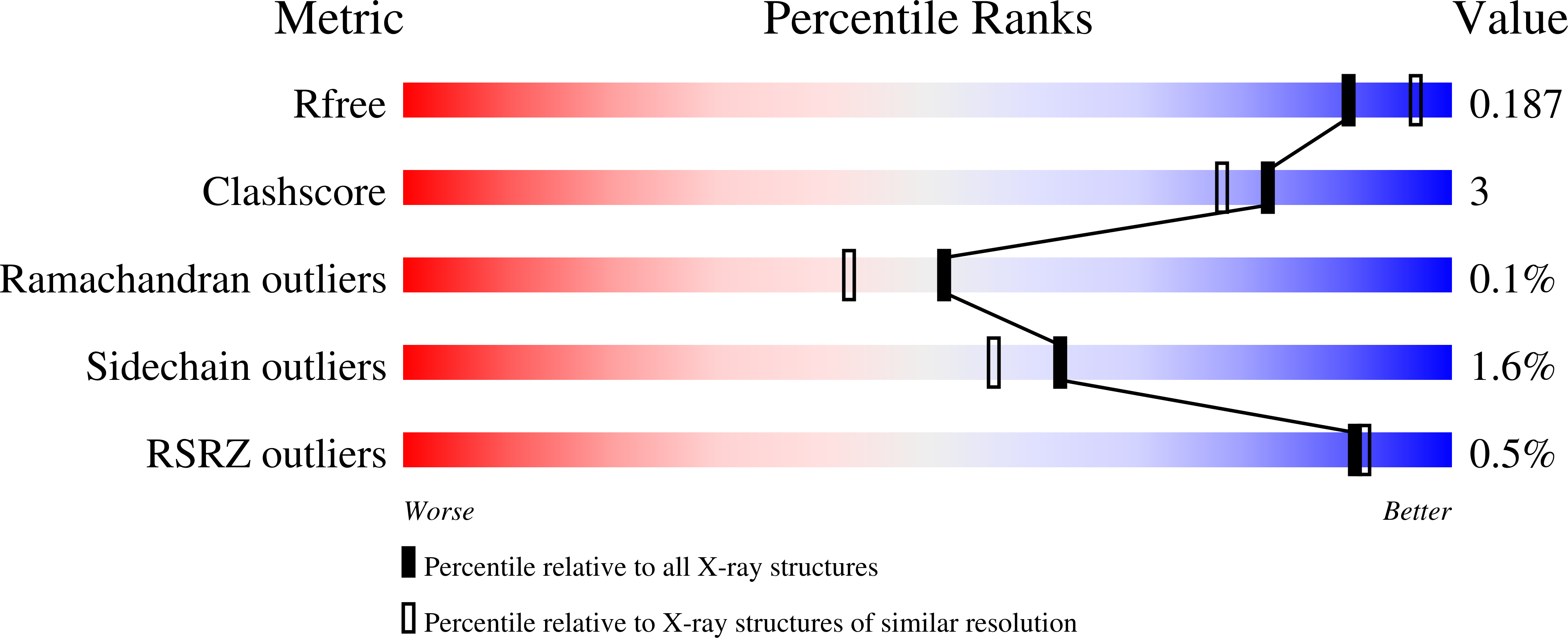
Resolution:
1.98 Å
R-Value Free:
0.18
R-Value Work:
0.15
R-Value Observed:
0.15
Space Group:
P 21 21 21