Deposition Date
2021-09-14
Release Date
2022-06-29
Last Version Date
2023-10-18
Entry Detail
PDB ID:
7S6H
Keywords:
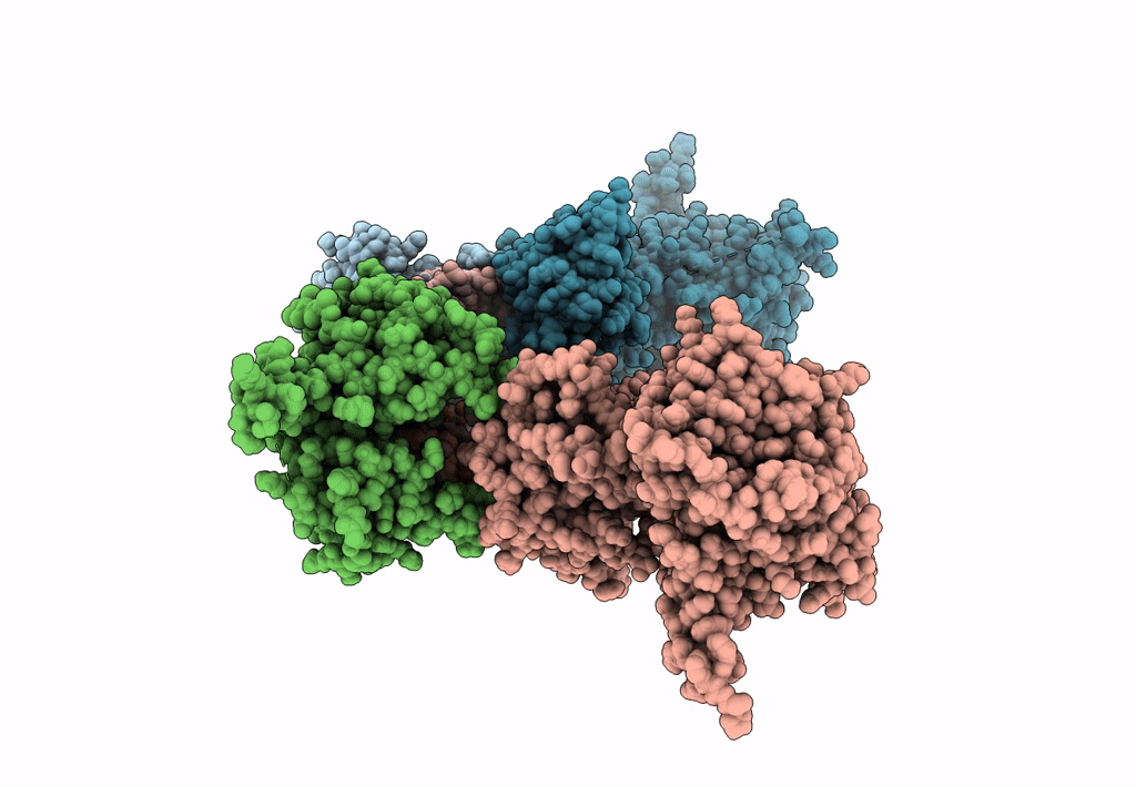
Title:
Human PARP1 deltaV687-E688 bound to NAD+ analog EB-47 and to a DNA double strand break.
Biological Source:
Source Organism(s):
Homo sapiens (Taxon ID: 9606)
synthetic construct (Taxon ID: 32630)
synthetic construct (Taxon ID: 32630)
Expression System(s):
Method Details:
Experimental Method:
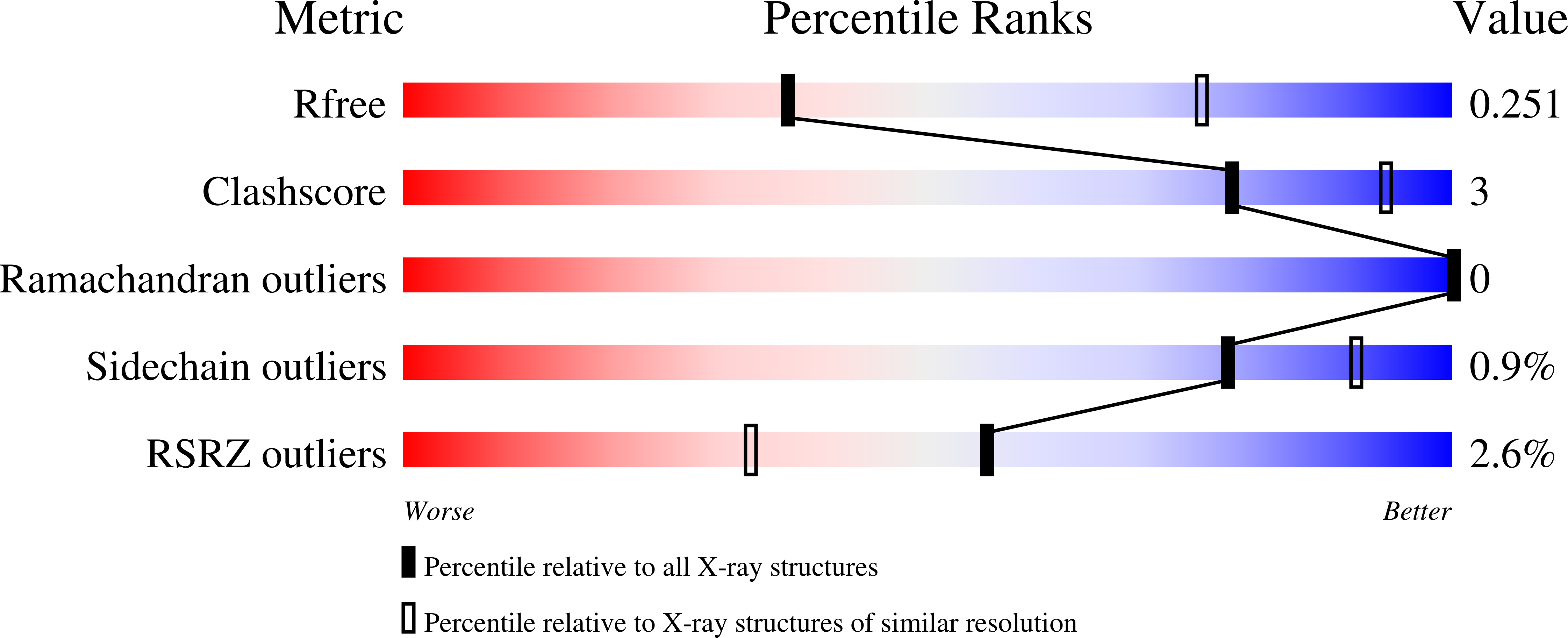
Resolution:
3.10 Å
R-Value Free:
0.25
R-Value Work:
0.21
R-Value Observed:
0.21
Space Group:
P 1 21 1