7S68 image
Deposition Date 2021-09-13
Release Date 2022-06-29
Last Version Date 2023-10-18
Entry Detail
PDB ID:
7S68
Title:
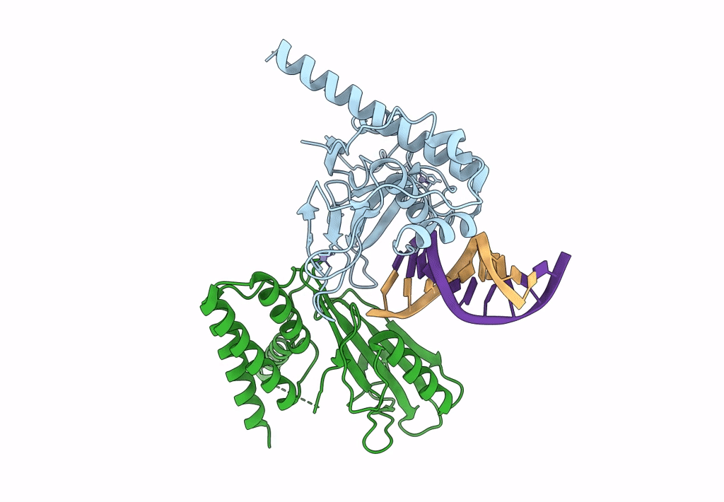
Structure of human PARP1 domains (Zn1, Zn3, WGR and HD) bound to a DNA double strand break.
Biological Source:
Source Organism:
Homo sapiens (Taxon ID: 9606)
DNA molecule (Taxon ID: 2853804)
Host Organism:
Method Details:
Experimental Method:
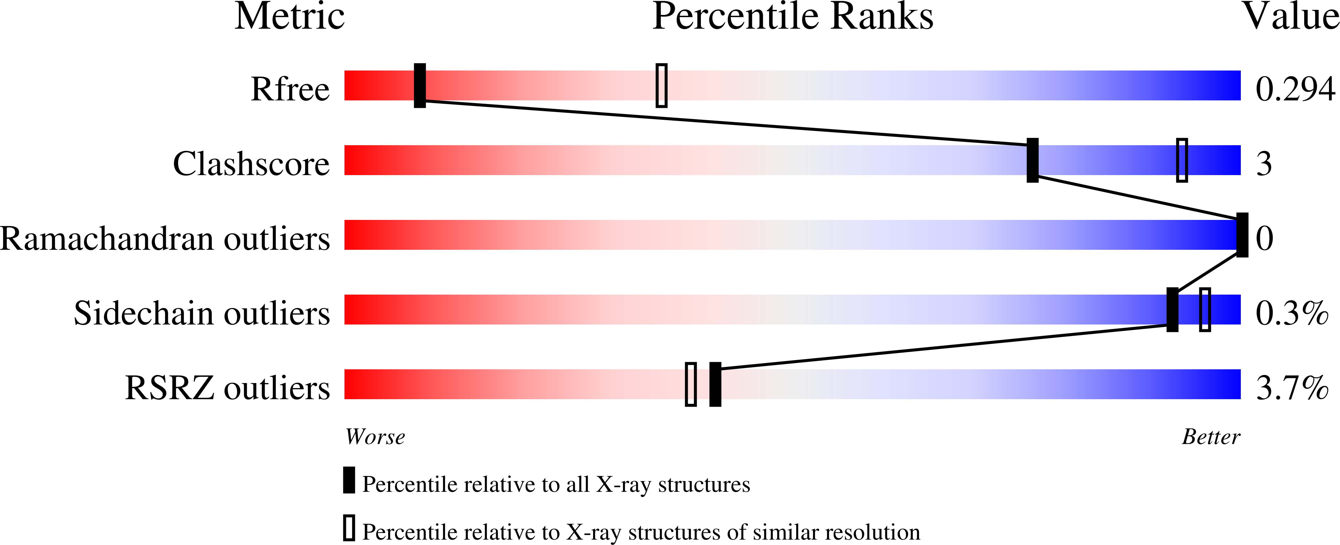
Resolution:
3.30 Å
R-Value Free:
0.29
R-Value Work:
0.25
R-Value Observed:
0.25
Space Group:
C 1 2 1
Macromolecular Entities
Structures with similar UniProt ID
Protein Blast
Polymer Type:polypeptide(L)
Molecule:Fusion of PARP1 zinc fingers 1 and 3 (Zn1, Zn3)
Gene (Uniprot):PARP1
Chain IDs:D (auth: A)
Chain Length:276
Number of Molecules:1
Biological Source:Homo sapiens
Structures with similar UniProt ID
Protein Blast
Polymer Type:polypeptide(L)
Molecule:Poly [ADP-ribose] polymerase 1
Gene (Uniprot):PARP1
Chain IDs:C (auth: B)
Chain Length:266
Number of Molecules:1
Biological Source:Homo sapiens
Polymer Type:polydeoxyribonucleotide
Molecule:DNA (5'-D(*GP*CP*CP*TP*GP*CP*AP*GP*GP*C)-3')
Chain IDs:A (auth: M), B (auth: N)
Chain Length:10
Number of Molecules:2
Biological Source:DNA molecule
Ligand Molecules
Primary Citation
Captured snapshots of PARP1 in the active state reveal the mechanics of PARP1 allostery.
Mol.Cell 82 2939 2951.e5 (2022)
PMID: 35793673 DOI: 10.1016/j.molcel.2022.06.011

Abstact

PARP1 rapidly detects DNA strand break damage and allosterically signals break detection to the PARP1 catalytic domain to activate poly(ADP-ribose) production from NAD+. PARP1 activation is characterized by dynamic changes in the structure of a regulatory helical domain (HD); yet, there are limited insights into the specific contributions that the HD makes to PARP1 allostery. Here, we have determined crystal structures of PARP1 in isolated active states that display specific HD conformations. These captured snapshots and biochemical analysis illustrate HD contributions to PARP1 multi-domain and high-affinity interaction with DNA damage, provide novel insights into the mechanics of PARP1 allostery, and indicate how HD active conformations correspond to alterations in the catalytic region that reveal the active site to NAD+. Our work deepens the understanding of PARP1 catalytic activation, the dynamics of the binding site of PARP inhibitor compounds, and the mechanisms regulating PARP1 retention on DNA damage.

Legend

Protein

Chemical

Disease

Primary Citation of related structures