7S5O image
Deposition Date 2021-09-11
Release Date 2022-03-30
Last Version Date 2024-10-23
Entry Detail
PDB ID:
7S5O
Title:
Crystal structure of Cytochrome c' beta from Nitrosomonas europaea ATCC 19718
Biological Source:
Method Details:
Experimental Method:
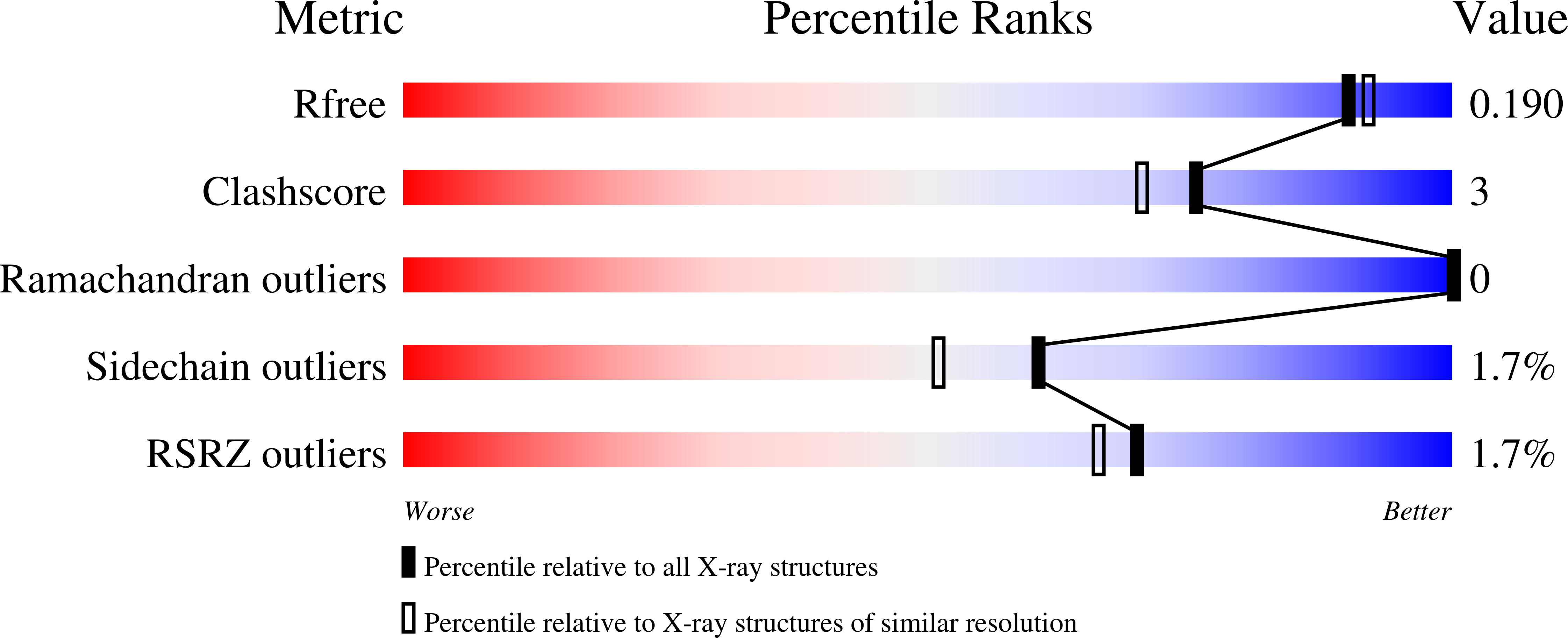
Resolution:
1.80 Å
R-Value Free:
0.19
R-Value Work:
0.15
R-Value Observed:
0.15
Space Group:
P 1 21 1
Macromolecular Entities
Structures with similar UniProt ID
Protein Blast
Polymer Type:polypeptide(L)
Molecule:Cytochrome_P460 domain-containing protein
Gene (Uniprot):NE0824
Chain IDs:A, B, C
Chain Length:169
Number of Molecules:3
Biological Source:Nitrosomonas europaea (strain ATCC 19718 / CIP 103999 / KCTC 2705 / NBRC 14298)
Primary Citation
Structural Characterization of Cytochrome c ' beta-Met from an Ammonia-Oxidizing Bacterium.
Biochemistry 61 563 574 (2022)
PMID: 35315646 DOI: 10.1021/acs.biochem.1c00640

Abstact

The ammonia-oxidizing bacterium Nitrosomonas europaea expresses two cytochromes in the P460 superfamily that are predicted to be structurally similar. In one, cytochrome (cyt) P460, the substrate hydroxylamine (NH2OH) is converted to nitric oxide (NO) and nitrous oxide (N2O) requiring a unique heme-lysyl cross-link in the catalytic cofactor. In the second, cyt c'β-Met, the cross-link is absent, and the cytochrome instead binds H2O2 forming a ferryl species similar to compound II of peroxidases. Here, we report the 1.80 Å crystal structure of cyt c'β-Met─a well-expressed protein in N. europaea with a lysine to a methionine replacement at the cross-linking position. The structure of cyt c'β-Met is characterized by a large β-sheet typical of P460 members; however, several localized structural differences render cyt c'β-Met distinct. This includes a large lasso-like loop at the "top" of the cytochrome that is not observed in other structurally characterized members. Active site variation is also observed, especially in comparison to its closest homologue cyt c'β from the methane-oxidizing Methylococcus capsulatus Bath, which also lacks the cross-link. The phenylalanine "cap" which is presumed to control small ligand access to the distal heme iron is replaced with an arginine, reminiscent of the strictly conserved distal arginine in peroxidases and to the NH2OH-oxidizing cytochromes P460. A critical proton-transferring glutamate residue required for NH2OH oxidation is nevertheless missing in the active site. This in part explains the inability of cyt c'β-Met to oxidize NH2OH. Our structure also rationalizes the absence of a methionyl cross-link, although the side chain's spatial position in the structure does not eliminate the possibility that it could form under certain conditions.

Legend

Protein

Chemical

Disease

Primary Citation of related structures
Feedback Form
Name
Email
Institute
Feedback