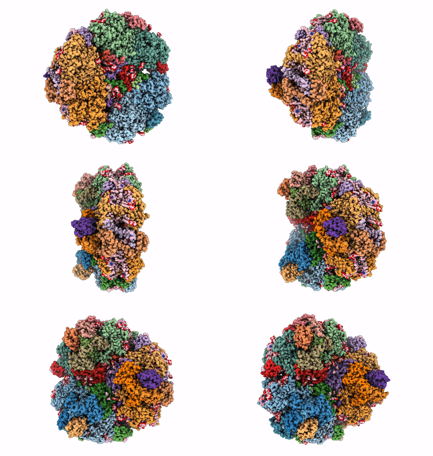
7S3D image
Deposition Date 2021-09-05
Release Date 2021-11-24
Last Version Date 2024-06-05
Entry Detail
PDB ID:
7S3D
Keywords:
Title:
Structure of photosystem I with bound ferredoxin from Synechococcus sp. PCC 7335 acclimated to far-red light
Biological Source:
Source Organism:
Method Details:
Experimental Method:
Resolution:
2.91 Å
Aggregation State:
PARTICLE
Reconstruction Method:
SINGLE PARTICLE
Macromolecular Entities
Polymer Type:polypeptide(L)
Molecule:Photosystem I P700 chlorophyll a apoprotein A1
Gene (Uniprot):psaA
Chain IDs:A, M (auth: G), Y (auth: a)
Chain Length:782
Number of Molecules:3
Biological Source:Synechococcus sp. PCC 7335
Polymer Type:polypeptide(L)
Molecule:Photosystem I P700 chlorophyll a apoprotein A2
Gene (Uniprot):psaB
Chain IDs:B, N (auth: H), Z (auth: b)
Chain Length:743
Number of Molecules:3
Biological Source:Synechococcus sp. PCC 7335
Polymer Type:polypeptide(L)
Molecule:PsaC
Chain IDs:C, O (auth: N), AA (auth: c)
Chain Length:81
Number of Molecules:3
Biological Source:Synechococcus sp. PCC 7335
Polymer Type:polypeptide(L)
Molecule:Photosystem I 16 kDa polypeptide
Gene (Uniprot):S7335_2287
Chain IDs:D, P (auth: O), BA (auth: d)
Chain Length:155
Number of Molecules:3
Biological Source:Synechococcus sp. PCC 7335
Polymer Type:polypeptide(L)
Molecule:Photosystem I reaction center subunit IV
Gene (Uniprot):psaE
Chain IDs:E, Q (auth: P), CA (auth: e)
Chain Length:71
Number of Molecules:3
Biological Source:Synechococcus sp. PCC 7335
Polymer Type:polypeptide(L)
Molecule:PSI-F
Gene (Uniprot):S7335_5139
Chain IDs:F, R (auth: Q), DA (auth: f)
Chain Length:168
Number of Molecules:3
Biological Source:Synechococcus sp. PCC 7335
Polymer Type:polypeptide(L)
Molecule:PsaI2
Gene (Uniprot):S7335_2282
Chain IDs:G (auth: I), S (auth: R), EA (auth: i)
Chain Length:70
Number of Molecules:3
Biological Source:Synechococcus sp. PCC 7335
Polymer Type:polypeptide(L)
Molecule:Photosystem I reaction center subunit IX
Gene (Uniprot):S7335_5198
Chain IDs:H (auth: J), T (auth: S), FA (auth: j)
Chain Length:46
Number of Molecules:3
Biological Source:Synechococcus sp. PCC 7335
Polymer Type:polypeptide(L)
Molecule:Photosystem I reaction center subunit PsaK
Gene (Uniprot):psaK
Chain IDs:I (auth: K), U (auth: T), GA (auth: k)
Chain Length:84
Number of Molecules:3
Biological Source:Synechococcus sp. PCC 7335
Polymer Type:polypeptide(L)
Molecule:PSI subunit V
Gene (Uniprot):S7335_2771
Chain IDs:J (auth: L), V (auth: U), HA (auth: l)
Chain Length:174
Number of Molecules:3
Biological Source:Synechococcus sp. PCC 7335
Polymer Type:polypeptide(L)
Molecule:PsaM
Chain IDs:K (auth: M), W (auth: V), IA (auth: m)
Chain Length:31
Number of Molecules:3
Biological Source:Synechococcus sp. PCC 7335
Polymer Type:polypeptide(L)
Molecule:2Fe-2S ferredoxin-type domain-containing protein
Gene (Uniprot):S7335_2822
Chain IDs:L (auth: X), X (auth: W), JA (auth: x)
Chain Length:99
Number of Molecules:3
Biological Source:Synechococcus sp. PCC 7335
Primary Citation
Structure of a photosystem I-ferredoxin complex from a marine cyanobacterium provides insights into far-red light photoacclimation.
J.Biol.Chem. 298 101408 101408 (2021)
PMID: 34793839 DOI: 10.1016/j.jbc.2021.101408

Abstact

Far-red light photoacclimation exhibited by some cyanobacteria allows these organisms to use the far-red region of the solar spectrum (700-800 nm) for photosynthesis. Part of this process includes the replacement of six photosystem I (PSI) subunits with isoforms that confer the binding of chlorophyll (Chl) f molecules that absorb far-red light (FRL). However, the exact sites at which Chl f molecules are bound are still challenging to determine. To aid in the identification of Chl f-binding sites, we solved the cryo-EM structure of PSI from far-red light-acclimated cells of the cyanobacterium Synechococcus sp. PCC 7335. We identified six sites that bind Chl f with high specificity and three additional sites that are likely to bind Chl f at lower specificity. All of these binding sites are in the core-antenna regions of PSI, and Chl f was not observed among the electron transfer cofactors. This structural analysis also reveals both conserved and nonconserved Chl f-binding sites, the latter of which exemplify the diversity in FRL-PSI among species. We found that the FRL-PSI structure also contains a bound soluble ferredoxin, PetF1, at low occupancy, which suggests that ferredoxin binds less transiently than expected according to the canonical view of ferredoxin-binding to facilitate electron transfer. We suggest that this may result from structural changes in FRL-PSI that occur specifically during FRL photoacclimation.

Legend

Protein

Chemical

Disease

Primary Citation of related structures