Deposition Date
2021-08-26
Release Date
2023-02-22
Last Version Date
2024-10-30
Entry Detail
PDB ID:
7RYT
Keywords:
Title:
Crystal structure of Mycobacterium tuberculosis acetylated Homoserine transacetylase with Coenzyme A
Biological Source:
Source Organism(s):
Mycobacterium tuberculosis (Taxon ID: 1773)
Expression System(s):
Method Details:
Experimental Method:
Resolution:
2.67 Å
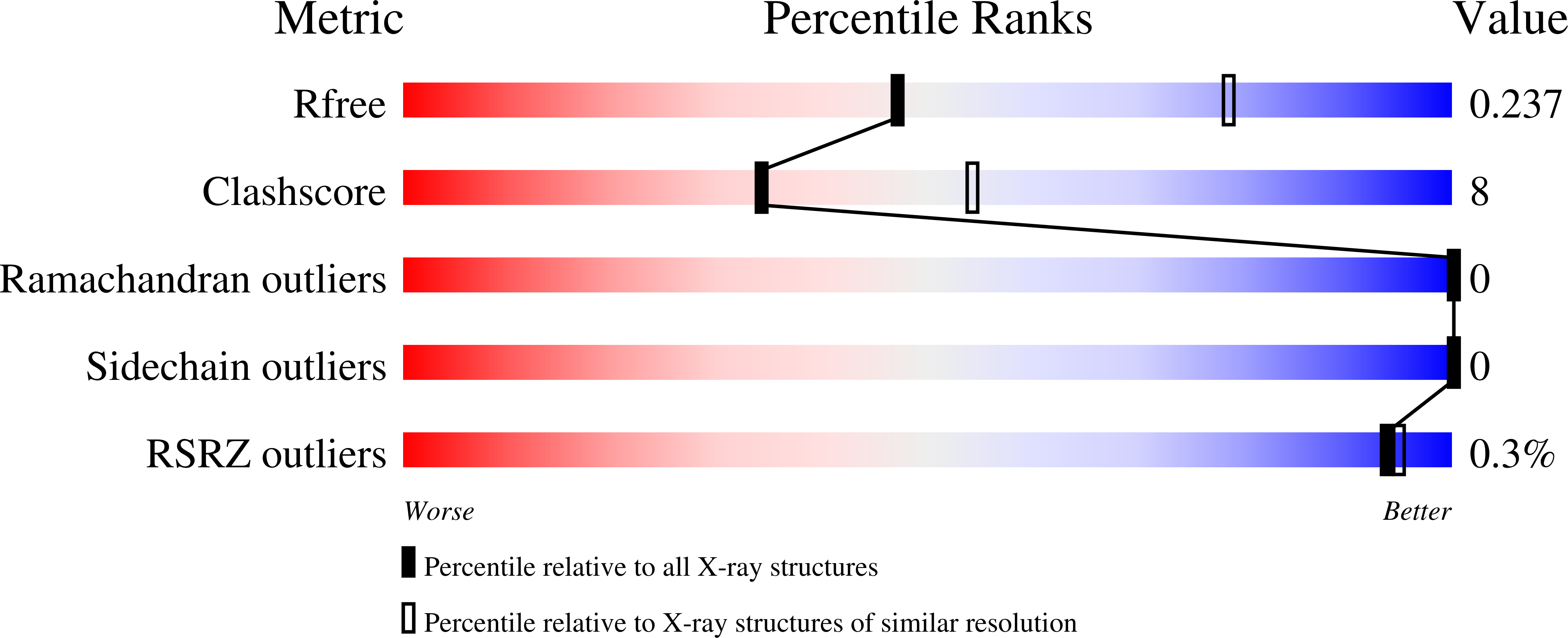
R-Value Free:
0.23
R-Value Work:
0.17
R-Value Observed:
0.18
Space Group:
P 21 21 21