7RXQ image
Deposition Date 2021-08-23
Release Date 2022-02-23
Last Version Date 2023-10-18
Entry Detail
PDB ID:
7RXQ
Title:
Crystal structure of junctophilin-2 in complex with a CaV1.1 peptide
Biological Source:
Source Organism:
Host Organism:
Method Details:
Experimental Method:
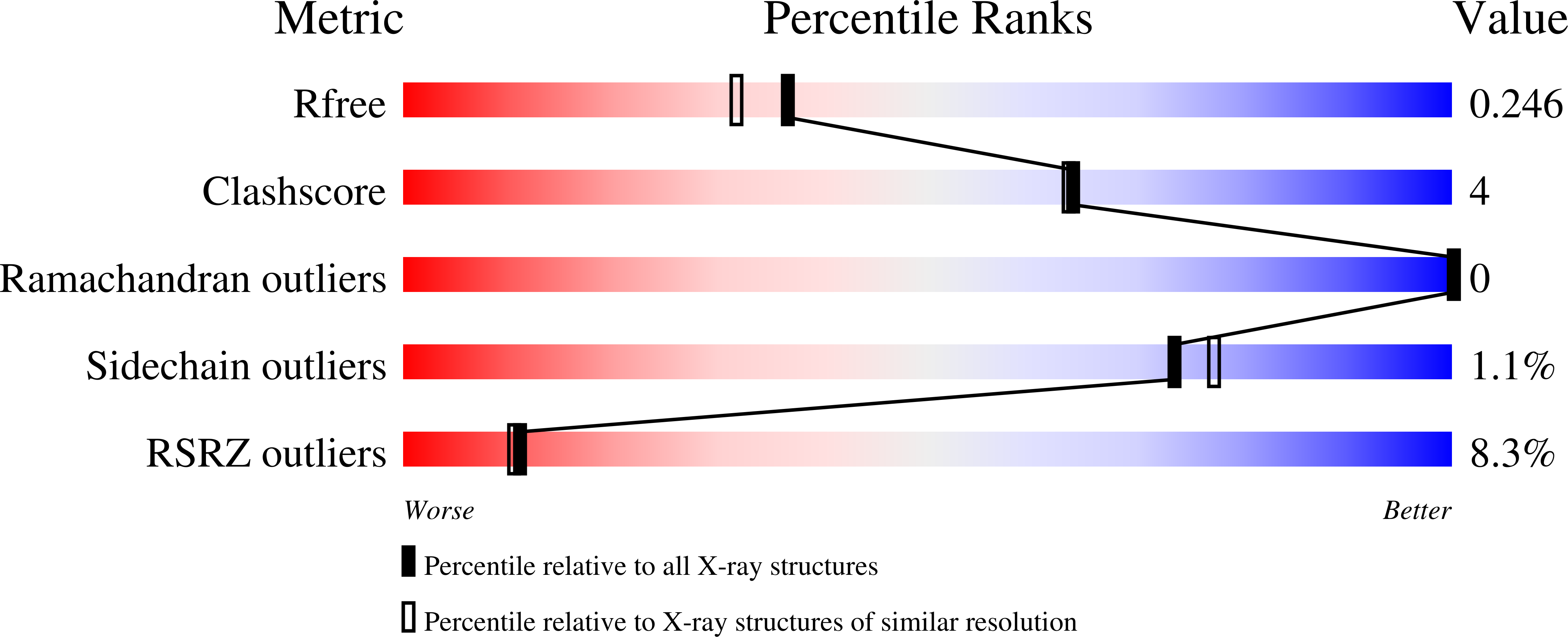
Resolution:
2.03 Å
R-Value Free:
0.25
R-Value Work:
0.20
R-Value Observed:
0.20
Space Group:
C 1 2 1
Macromolecular Entities
Polymer Type:polypeptide(L)
Molecule:Junctophilin-2 N-terminal fragment
Gene (Uniprot):JPH2
Chain IDs:A
Chain Length:327
Number of Molecules:1
Biological Source:Homo sapiens
Polymer Type:polypeptide(L)
Molecule:Voltage-dependent L-type calcium channel subunit alpha-1S
Gene (Uniprot):CACNA1S
Chain IDs:B
Chain Length:16
Number of Molecules:1
Biological Source:Oryctolagus cuniculus
Primary Citation
Structures of the junctophilin/voltage-gated calcium channel interface reveal hot spot for cardiomyopathy mutations.
Proc.Natl.Acad.Sci.USA 119 e2120416119 e2120416119 (2022)
PMID: 35238659 DOI: 10.1073/pnas.2120416119

Abstact

SignificanceIon channels have evolved the ability to communicate with one another, either through protein-protein interactions, or indirectly via intermediate diffusible messenger molecules. In special cases, the channels are part of different membranes. In muscle tissue, the T-tubule membrane is in proximity to the sarcoplasmic reticulum, allowing communication between L-type calcium channels and ryanodine receptors. This process is critical for excitation-contraction coupling and requires auxiliary proteins like junctophilin (JPH). JPHs are targets for disease-associated mutations, most notably hypertrophic cardiomyopathy mutations in the JPH2 isoform. Here we provide high-resolution snapshots of JPH, both alone and in complex with a calcium channel peptide, and show how this interaction is targeted by cardiomyopathy mutations.

Legend

Protein

Chemical

Disease

Primary Citation of related structures