Deposition Date
2021-08-11
Release Date
2022-07-20
Last Version Date
2024-05-22
Entry Detail
Biological Source:
Source Organism(s):
Rotavirus B (isolate RVB/Human/China/ADRV/1982) (Taxon ID: 10942)
Rotavirus (Taxon ID: 10912)
Rotavirus (Taxon ID: 10912)
Expression System(s):
Method Details:
Experimental Method:
Resolution:
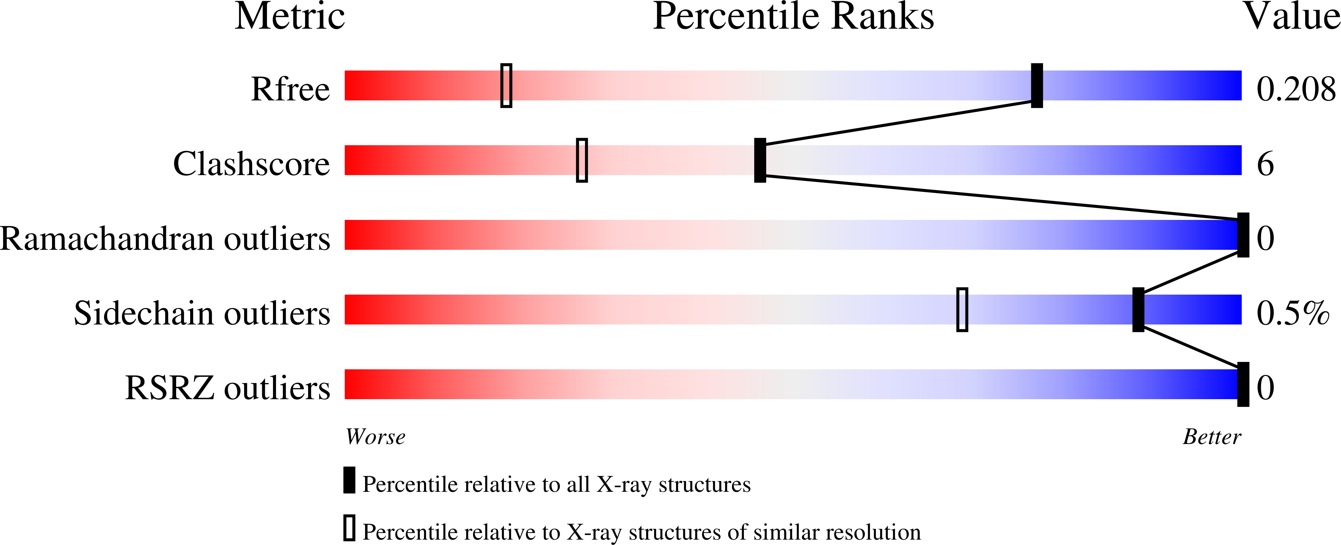
1.32 Å
R-Value Free:
0.20
R-Value Work:
0.15
R-Value Observed:
0.15
Space Group:
P 1 21 1