7RS1 image
Entry Detail
PDB ID:
7RS1
Title:
Crystal Structure of the ER-alpha Ligand-binding Domain (L372S, L536S) in complex with DMERI-21
Biological Source:
Source Organism:
PDB Version:
Deposition Date:
2021-08-10
Release Date:
2021-09-08
Method Details:
Experimental Method:
Resolution:
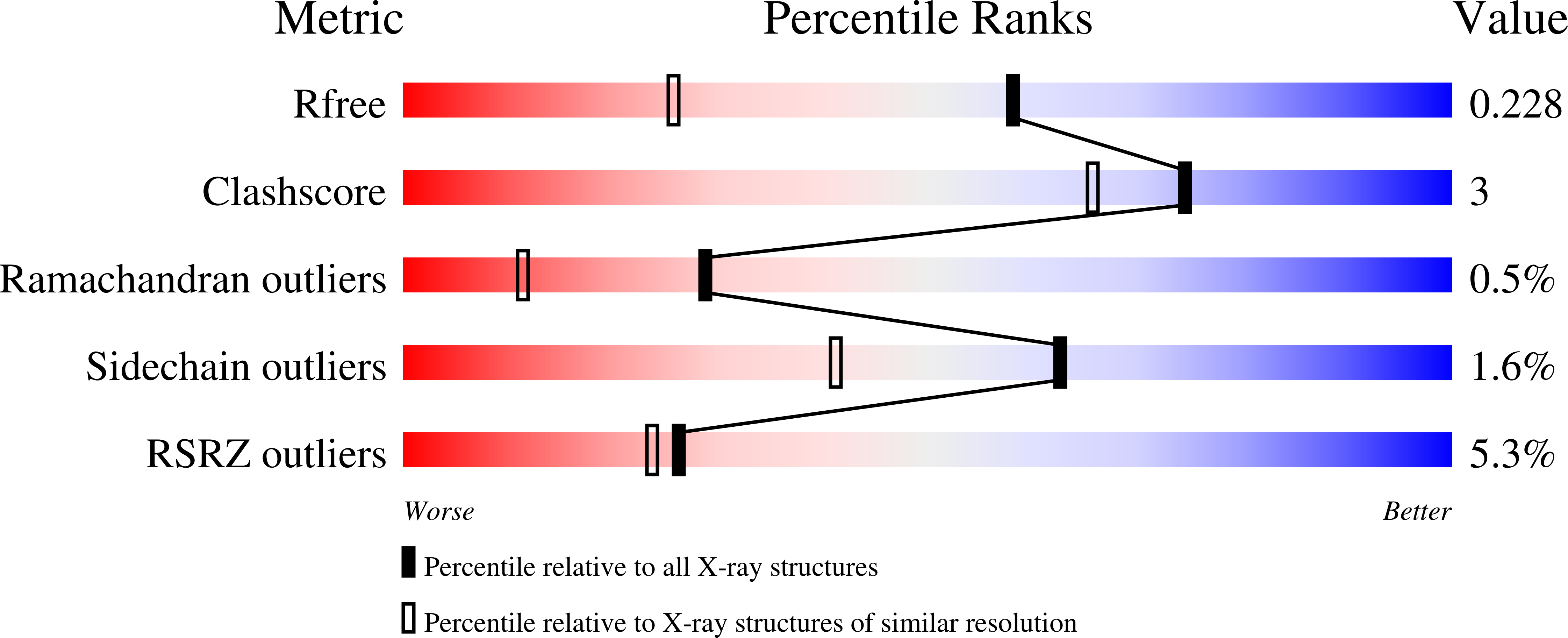
1.59 Å
R-Value Free:
0.21
R-Value Work:
0.18
R-Value Observed:
0.18
Space Group:
P 1
Macromolecular Entities
Polymer Type:polypeptide(L)
Description:Estrogen receptor
Mutations:L372S, L536S
Chain IDs:A, B, C, D
Chain Length:257
Number of Molecules:4
Biological Source:Homo sapiens
Modified Residue
Compound ID Chain ID Parent Comp ID Details 2D Image
YCM A CYS modified residue
Ligand Molecules
Primary Citation

Abstact

Efforts to improve estrogen receptor-α (ER)-targeted therapies in breast cancer have relied upon a single mechanism, with ligands having a single side chain on the ligand core that extends outward to determine antagonism of breast cancer growth. Here, we describe inhibitors with two ER-targeting moieties, one of which uses an alternate structural mechanism to generate full antagonism, freeing the side chain to independently determine other critical properties of the ligands. By combining two molecular targeting approaches into a single ER ligand, we have generated antiestrogens that function through new mechanisms and structural paradigms to achieve antagonism. These dual-mechanism ER inhibitors (DMERIs) cause alternate, noncanonical structural perturbations of the receptor ligand-binding domain (LBD) to antagonize proliferation in ER-positive breast cancer cells and in allele-specific resistance models. Our structural analyses with DMERIs highlight marked differences from current standard-of-care, single-mechanism antiestrogens. These findings uncover an enhanced flexibility of the ER LBD through which it can access nonconsensus conformational modes in response to DMERI binding, broadly and effectively suppressing ER activity.

Legend

Protein

Chemical

Disease

Primary Citation of related structures