7RM5 image
Deposition Date 2021-07-26
Release Date 2021-09-08
Last Version Date 2024-11-20
Entry Detail
PDB ID:
7RM5
Title:
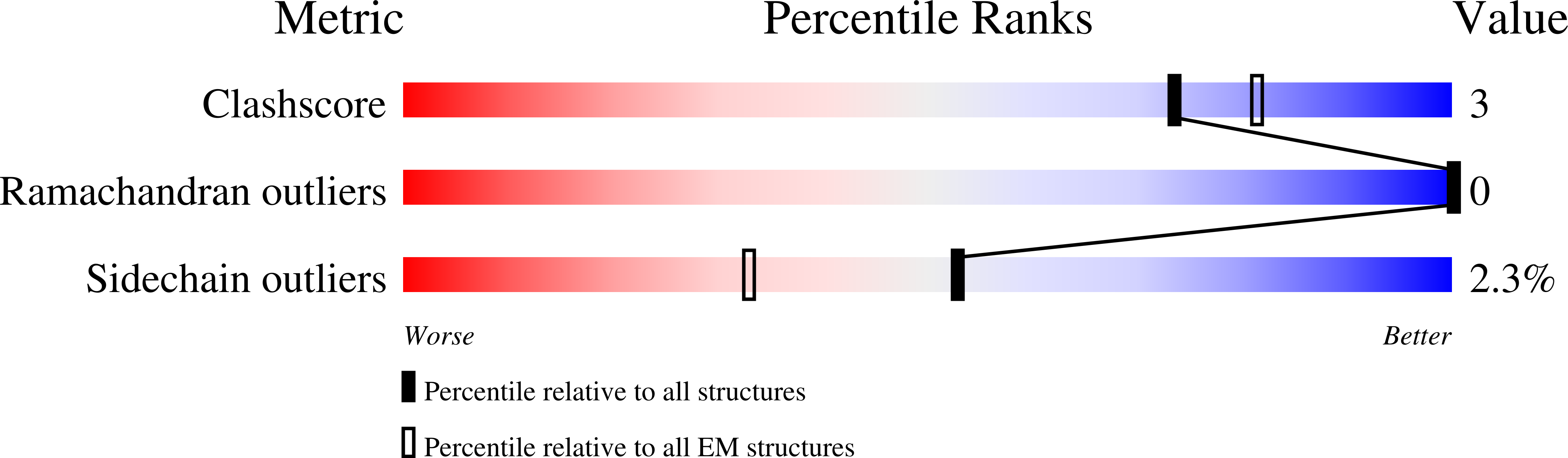
MicroED structure of the human adenosine receptor at 2.8A
Biological Source:
Source Organism:
Homo sapiens (Taxon ID: 9606)
Escherichia coli (Taxon ID: 562)
Host Organism:
Method Details:
Experimental Method:
Resolution:
2.79 Å
R-Value Free:
0.28
R-Value Work:
0.24
R-Value Observed:
0.25
Space Group:
C 2 2 21
Macromolecular Entities
Protein Blast
Polymer Type:polypeptide(L)
Molecule:Adenosine receptor A2a/Soluble cytochrome b562 chimera
Gene (Uniprot):cybC, ADORA2A
Chain IDs:A
Chain Length:447
Number of Molecules:1
Biological Source:Homo sapiens, Escherichia coli
Primary Citation
MicroED structure of the human adenosine receptor determined from a single nanocrystal in LCP.
Proc.Natl.Acad.Sci.USA 118 ? ? (2021)
PMID: 34462357 DOI: 10.1073/pnas.2106041118

Abstact

G protein-coupled receptors (GPCRs), or seven-transmembrane receptors, are a superfamily of membrane proteins that are critically important to physiological processes in the human body. Determining high-resolution structures of GPCRs without bound cognate signaling partners, such as a G protein, requires crystallization in lipidic cubic phase (LCP). GPCR crystals grown in LCP are often too small for traditional X-ray crystallography. These microcrystals are ideal for investigation by microcrystal electron diffraction (MicroED), but the gel-like nature of LCP makes traditional approaches to MicroED sample preparation insurmountable. Here, we show that the structure of a human A2A adenosine receptor can be determined by MicroED after converting the LCP into the sponge phase followed by focused ion-beam milling. We determined the structure of the A2A adenosine receptor to 2.8-Å resolution and resolved an antagonist in its orthosteric ligand-binding site, as well as four cholesterol molecules bound around the receptor. This study lays the groundwork for future structural studies of lipid-embedded membrane proteins by MicroED using single microcrystals that would be impossible with other crystallographic methods.

Legend

Protein

Chemical

Disease

Primary Citation of related structures
Feedback Form
Name
Email
Institute
Feedback