7REN image
Deposition Date 2021-07-13
Release Date 2022-05-25
Last Version Date 2023-10-18
Entry Detail
PDB ID:
7REN
Title:
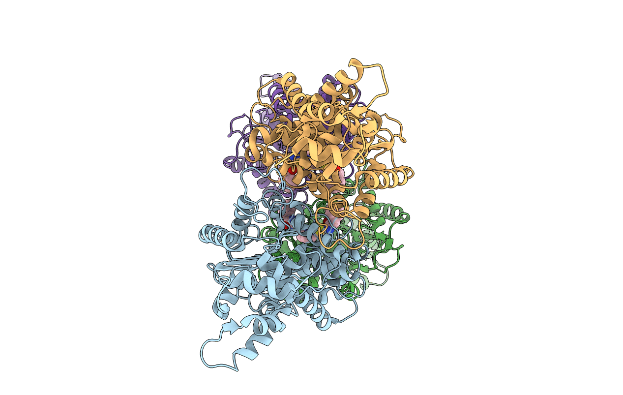
Room temperature serial crystal structure of Glutaminase C in complex with inhibitor UPGL-00004
Biological Source:
Source Organism:
Homo sapiens (Taxon ID: 9606)
Host Organism:
Method Details:
Experimental Method:
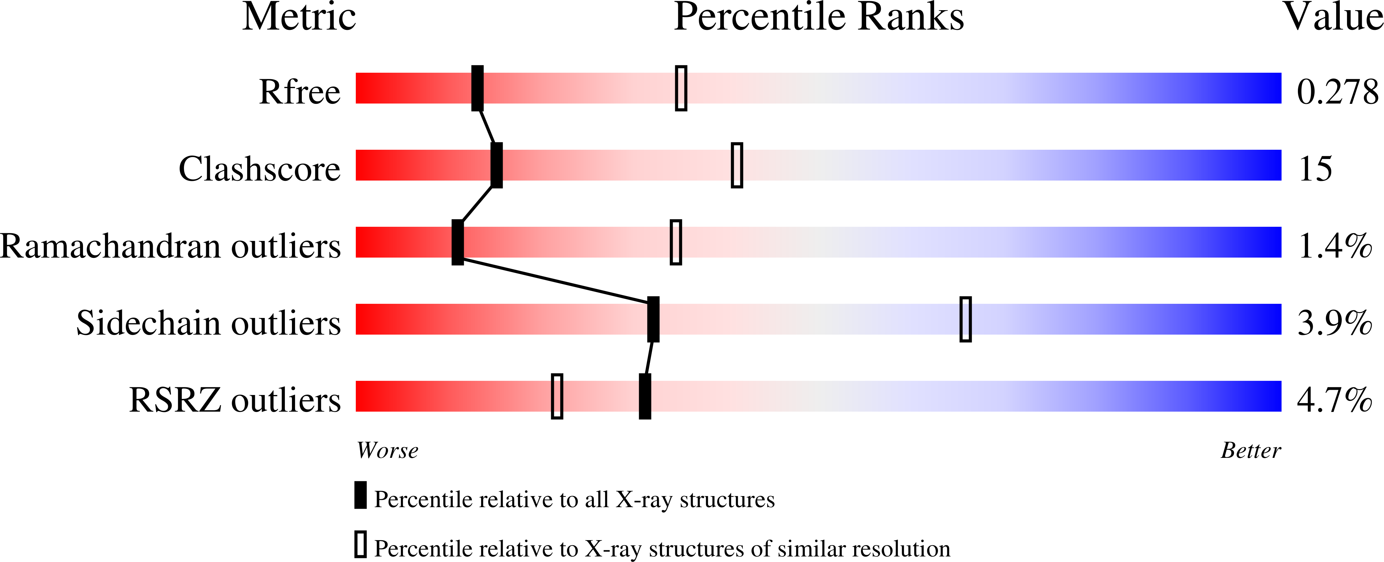
Resolution:
2.80 Å
R-Value Free:
0.27
R-Value Work:
0.20
R-Value Observed:
0.21
Space Group:
P 1 21 1
Macromolecular Entities
Polymer Type:polypeptide(L)
Molecule:Glutaminase kidney isoform, mitochondrial
Gene (Uniprot):GLS
Chain IDs:A, B, C, D
Chain Length:539
Number of Molecules:4
Biological Source:Homo sapiens
Ligand Molecules
Primary Citation
New insights into the molecular mechanisms of glutaminase C inhibitors in cancer cells using serial room temperature crystallography.
J.Biol.Chem. 298 101535 101535 (2022)
PMID: 34954143 DOI: 10.1016/j.jbc.2021.101535

Abstact

Cancer cells frequently exhibit uncoupling of the glycolytic pathway from the TCA cycle (i.e., the "Warburg effect") and as a result, often become dependent on their ability to increase glutamine catabolism. The mitochondrial enzyme Glutaminase C (GAC) helps to satisfy this 'glutamine addiction' of cancer cells by catalyzing the hydrolysis of glutamine to glutamate, which is then converted to the TCA-cycle intermediate α-ketoglutarate. This makes GAC an intriguing drug target and spurred the molecules derived from bis-2-(5-phenylacetamido-1,3,4-thiadiazol-2-yl)ethyl sulfide (the so-called BPTES class of allosteric GAC inhibitors), including CB-839, which is currently in clinical trials. However, none of the drugs targeting GAC are yet approved for cancer treatment and their mechanism of action is not well understood. Here, we shed new light on the underlying basis for the differential potencies exhibited by members of the BPTES/CB-839 family of compounds, which could not previously be explained with standard cryo-cooled X-ray crystal structures of GAC bound to CB-839 or its analogs. Using an emerging technique known as serial room temperature crystallography, we were able to observe clear differences between the binding conformations of inhibitors with significantly different potencies. We also developed a computational model to further elucidate the molecular basis of differential inhibitor potency. We then corroborated the results from our modeling efforts using recently established fluorescence assays that directly read out inhibitor binding to GAC. Together, these findings should aid in future design of more potent GAC inhibitors with better clinical outlook.

Legend

Protein

Chemical

Disease

Primary Citation of related structures