7RDF image
Deposition Date 2021-07-09
Release Date 2021-12-22
Last Version Date 2023-10-18
Entry Detail
PDB ID:
7RDF
Keywords:
Title:
Crystal structure of Pseudomonas aeruginosa D-Arginine Dehydrogenase Y249F co-crystallized in the presence of D-arginine
Biological Source:
Method Details:
Experimental Method:
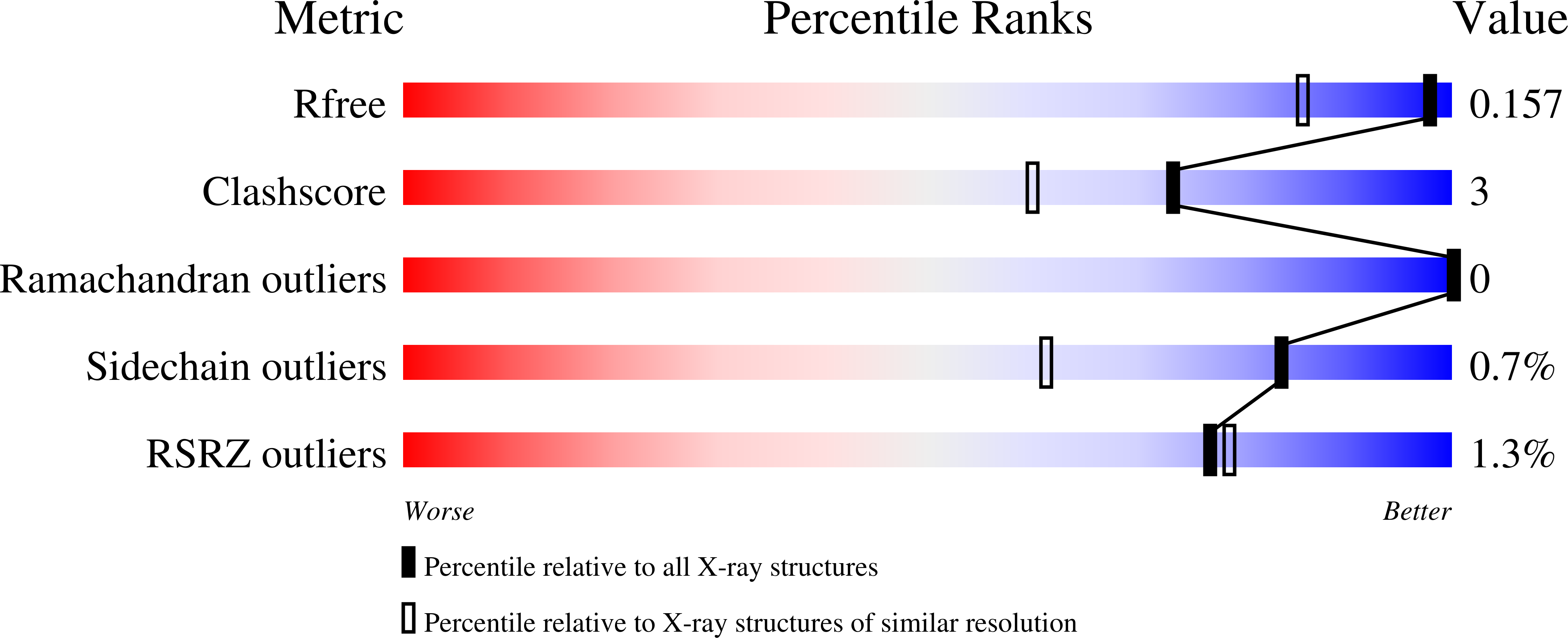
Resolution:
1.29 Å
R-Value Free:
0.15
R-Value Work:
0.12
R-Value Observed:
0.12
Space Group:
P 21 21 21
Macromolecular Entities
Structures with similar UniProt ID
Protein Blast
Polymer Type:polypeptide(L)
Molecule:FAD-dependent catabolic D-arginine dehydrogenase DauA
Gene (Uniprot):dauA
Chain IDs:A
Chain Length:375
Number of Molecules:1
Biological Source:Pseudomonas aeruginosa (strain ATCC 15692 / DSM 22644 / CIP 104116 / JCM 14847 / LMG 12228 / 1C / PRS 101 / PAO1)
Primary Citation
Discovery of a new flavin N5-adduct in a tyrosine to phenylalanine variant of d-Arginine dehydrogenase.
Arch.Biochem.Biophys. 715 109100 109100 (2021)
PMID: 34864048 DOI: 10.1016/j.abb.2021.109100

Abstact

d-Arginine dehydrogenase from Pseudomonas aeruginosa (PaDADH) catalyzes the flavin-dependent oxidation of d-arginine and other d-amino acids. Here, we report the crystal structure at 1.29 Å resolution for PaDADH-Y249F expressed and co-crystallized with d-arginine. The overall structure of PaDADH-Y249F resembled PaDADH-WT, but the electron density for the flavin cofactor was ambiguous, suggesting the presence of modified flavins. Electron density maps and mass spectrometric analysis confirmed the presence of both N5-(4-guanidino-oxobutyl)-FAD and 6-OH-FAD in a single crystal of PaDADH-Y249F and helped with the further refinement of the X-ray crystal structure. The versatility of the reduced flavin is apparent in the PaDADH-Y249F structure and is evidenced by the multiple functions it can perform in the same active site.

Legend

Protein

Chemical

Disease

Primary Citation of related structures
Feedback Form
Name
Email
Institute
Feedback